Multi-layer image
Description:
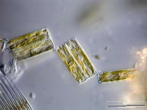
Colony of Diatoma vulgare. The mucilaginous connection material is shown. Scale bar indicates 50 µm.
Sample from Lake Constance near Bodman. Images were taken using Zeiss Universal with Olympus C7070 CCD camera.
Image under Creative Commons License V 3.0 (CC BY-NC-SA).
Included On The Following Pages:
- Life (creatures)
- Cellular (cellular organisms)
- Eukaryota (eukaryotes)
- SAR (Stramenopiles, Alveolates, Rhizaria)
- Stramenopiles (heterokont)
- Ochrophyta (Ochrophyte)
- Bacillariophyta (diatoms)
- Fragilariophyceae
- Fragilariophycidae
- Fragilariales
- Fragilariaceae
- Diatoma
- Diatoma vulgaris
- Oomycota (oomycetes)
- Diatomista
This image is not featured in any collections.
Source Information
- license
- cc-by-nc
- provider
- micro*scope
- original
- original media file
- visit source
- partner site
- micro*scope
- ID