in vivo;lRight lateral view
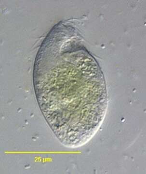
Description:
Right lateral view of the colorles ciliate Trimyema compressum (Lackey,1925).The cell is fusiform with bluntly rounded anterior and posterior ends. The funnel-shaped anterior oral aperture is subapical (seen here). 50-60 somatic kineties are reduced to three cilated basal bodies in each row.The arrangement of the somatic kineties gives the appearance of three discontinuous slightly spiral rows of cilia when viewed from ventral aspect. There is a single long caudal cilium (not seen here).Two long C- shaped kineties border the oral aperture. There is a short 3rd innermost kinety at the posterior end of the oral aperture. Near the anterior end of the oral kineties is a smal group of dikinetids representing the "adoral membranelles". There is a spherical central macronucleus.A single lateral contractile vacuole is located in the posterior 1/2 of the cell.From polysaprobic sediments of a freshwater rain barrel near Boise, Idaho.December 2005.DIC.
Included On The Following Pages:
- Life (creatures)
- Cellular (cellular organisms)
- Eukaryota (eukaryotes)
- SAR (Stramenopiles, Alveolates, Rhizaria)
- Alveolata (alveolates)
- Ciliophora (ciliates)
- Intramacronucleata
- Litostomatea
- Trichostomatia
- Vestibuliferida
- Trimyemidae
- Trimyema
- Trimyema compressum
This image is not featured in any collections.
Source Information
- license
- cc-by-nc
- author
- William Bourland
- provider
- micro*scope
- original
- original media file
- visit source
- partner site
- micro*scope
- ID