Multilayer image (DOF)
Description:
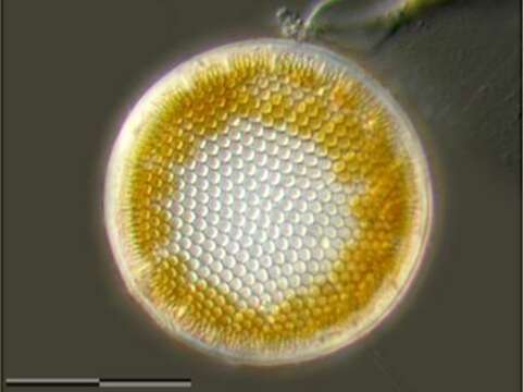
Marginal silica processes are visible. Scale bar indicates 25 µm. The image was built up using several photomicrographic frames with manual stacking technique.
Sample from North Sea near Heligoland (spring diatom bloom). Images were taken using Zeiss Universal with Olympus C7070 CCD camera.
Included On The Following Pages:
- Life (creatures)
- Cellular (cellular organisms)
- Eukaryota (eukaryotes)
- SAR (Stramenopiles, Alveolates, Rhizaria)
- Stramenopiles (heterokont)
- Oomycota (oomycetes)
- Ochrophyta (Ochrophyte)
- Diatomista
- Bacillariophyta (diatoms)
- Coscinodiscophyceae
- Thalassiosirophycidae
- Thalassiosirales
- Thalassiosiraceae
- Thalassiosira
This image is not featured in any collections.
Source Information
- license
- cc-by-nc
- provider
- micro*scope
- original
- original media file
- visit source
- partner site
- micro*scope
- ID