portrait
Description:
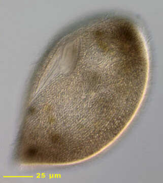
Portrait of the widely distributed hymenostome ciliate, Frontonia atra. This species has a distinctive dorsoventrally flattened teardrop shape. The anterior is broadly rounded, the posterior tapering to a blunt point. F. atra may be confused with Disematostoma buetschlii (the latter has a distinctive cross-striated pre and postoral ciliary suture and contains zoochlorellae and/or kleptoplasts). F. atra has a dense aggregate of dark brown cytoplasmic granules anteriorly (possibly endosymbiotic bacteria). The oral aperture is roughly triangular with the base posterior and the anterior apex terminating at a thin preoral suture. There is an undulating membrane on the right and three adoral membranelles on the left. There is a narrow postoral suture to the right of which lie prominent vestibular ciliary rows and to the left of which lie postoral kineties. The round macronucleus is seen here anterior and to the left of the oral aperture. Numerous extrusomes form a peripheral fringe. The single contractile vacuole (not seen here) is subequatorial on the right. Probably omnivorous. Often found feeding on diatoms and green algae. Collected from freshwater pond near Boise, Idaho in June 2003. DIC optics.
Included On The Following Pages:
- Life (creatures)
- Cellular (cellular organisms)
- Eukaryota (eukaryotes)
- SAR (Stramenopiles, Alveolates, Rhizaria)
- Alveolata (alveolates)
- Ciliophora (ciliates)
- Intramacronucleata
- Oligohymenophorea
- Peniculida (Peniculid)
- Frontoniidae
- Frontonia
- Frontonia atra
This image is not featured in any collections.
Source Information
- license
- cc-by-nc
- author
- William Bourland
- provider
- micro*scope
- original
- original media file
- visit source
- partner site
- micro*scope
- ID