Image of Austrarchaea aleenae Rix & Harvey 2011

Description:
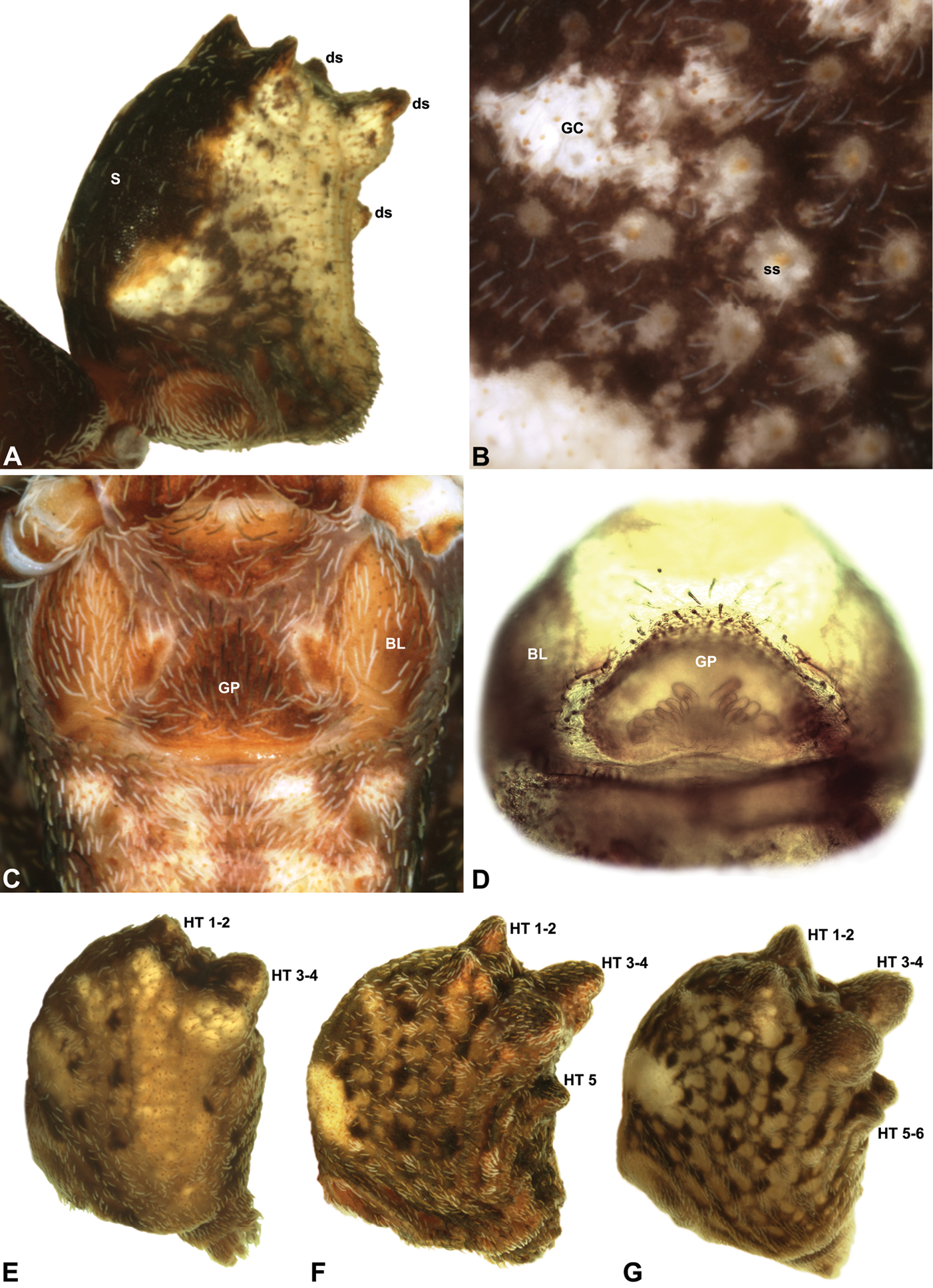
Figure 5.Abdominal morphology of Austrarchaea species. A–C, A. judyae sp. n.: A, male abdomen, antero-lateral view, showing dorsal scute (S) and additional dorsal sclerites (ds); B, detail of female abdomen, lateral view, showing subcuticular guanine crystals (GC) and concentric arrangements of setae around sclerotic spots (ss); C, female epigastric region, ventral view, showing setose book lung covers (BL) and genital plate (GP). D, Cleared epigastric region of female A. nodosa (Forster), postero-ventral view, showing position of clustered spermathecae under posterior rim of genital plate. E–G, Female abdomens, postero-lateral view, showing arrangement of dorsal hump-like tubercles (HT) in different taxa: E, A. sp. nr. daviesae (QMB S72989, from Mount Bartle Frere, NE. Queensland); F, A. monteithi sp. n.; G, A. aleenae sp. n. Note the presence of only a single posterior hump-like tubercle (HT 5) in A. monteithi.
Included On The Following Pages:
- Life (creatures)
- Cellular (cellular organisms)
- Eukaryota (eukaryotes)
- Opisthokonta (opisthokonts)
- Metazoa (Animal)
- Bilateria
- Protostomia (protostomes)
- Ecdysozoa (ecdysozoans)
- Arthropoda (arthropods)
- Chelicerata (chelicerates)
- Arachnida (arachnids)
- Araneae (spiders)
- Opisthothelae
- Araneomorphae
- Entelegynae
- Archaeidae (pelican spiders)
- Austrarchaea
- Austrarchaea aleenae
- Panarthropoda
This image is not featured in any collections.
Source Information
- license
- cc-by-3.0
- copyright
- Michael G. Rix, Mark S. Harvey
- bibliographic citation
- Rix M, Harvey M (2011) Australian Assassins, Part I: A review of the Assassin Spiders (Araneae, Archaeidae) of mid-eastern Australia ZooKeys 123: 1–100
- original
- original media file
- visit source
- partner site
- Zookeys
- ID