Image of Pselaphodes
Description:
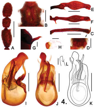
Figure 4. Details of male Pselaphodes shii. A left antennal club B pronotum C protibia D apex of protibia, enlarged E mesotrochanter and mesofemur F metatrochanter and metafemur G metaventral process, in lateral view H sternite IX I aedeagus, in lateral view J–K same, in dorsal view L right paramere, enlarged. Scales: A, B, C, E, F = 0.3 mm, D = 0.05 mm, G, H = 0.1 mm, L = 0.025 mm, I, J, K = 0.2 mm.
Included On The Following Pages:
- Life (creatures)
- Cellular (cellular organisms)
- Eukaryota (eukaryotes)
- Opisthokonta (opisthokonts)
- Metazoa (Animal)
- Bilateria
- Protostomia (protostomes)
- Ecdysozoa (ecdysozoans)
- Arthropoda (arthropods)
- Pancrustacea
- Hexapoda (hexapods)
- Insecta (insects)
- Pterygota (winged insects)
- Neoptera (neopteran)
- Endopterygota (endopterygotes)
- Coleoptera (beetles)
- Polyphaga
- Staphyliniformia
- Staphylinoidea
- Staphylinidae (rove beetles)
- Pselaphodes
- Pselaphodes shii
- Panarthropoda
This image is not featured in any collections.
Source Information
- license
- cc-by-3.0
- copyright
- Zi-Wei Yin, Li-Zhen Li, Mei-Jun Zhao
- bibliographic citation
- Yin Z, Li L, Zhao M (2012) Two new species of Pselaphodes Westwood and new record of Taiwanophodes minor Hlaváč from South China (Coleoptera, Staphylinidae, Pselaphinae) ZooKeys 175: 75–86
- original
- original media file
- visit source
- partner site
- Zookeys
- ID