Image of Sinopoda
Description:
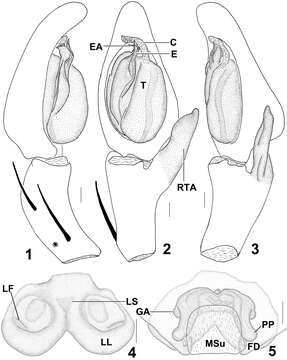
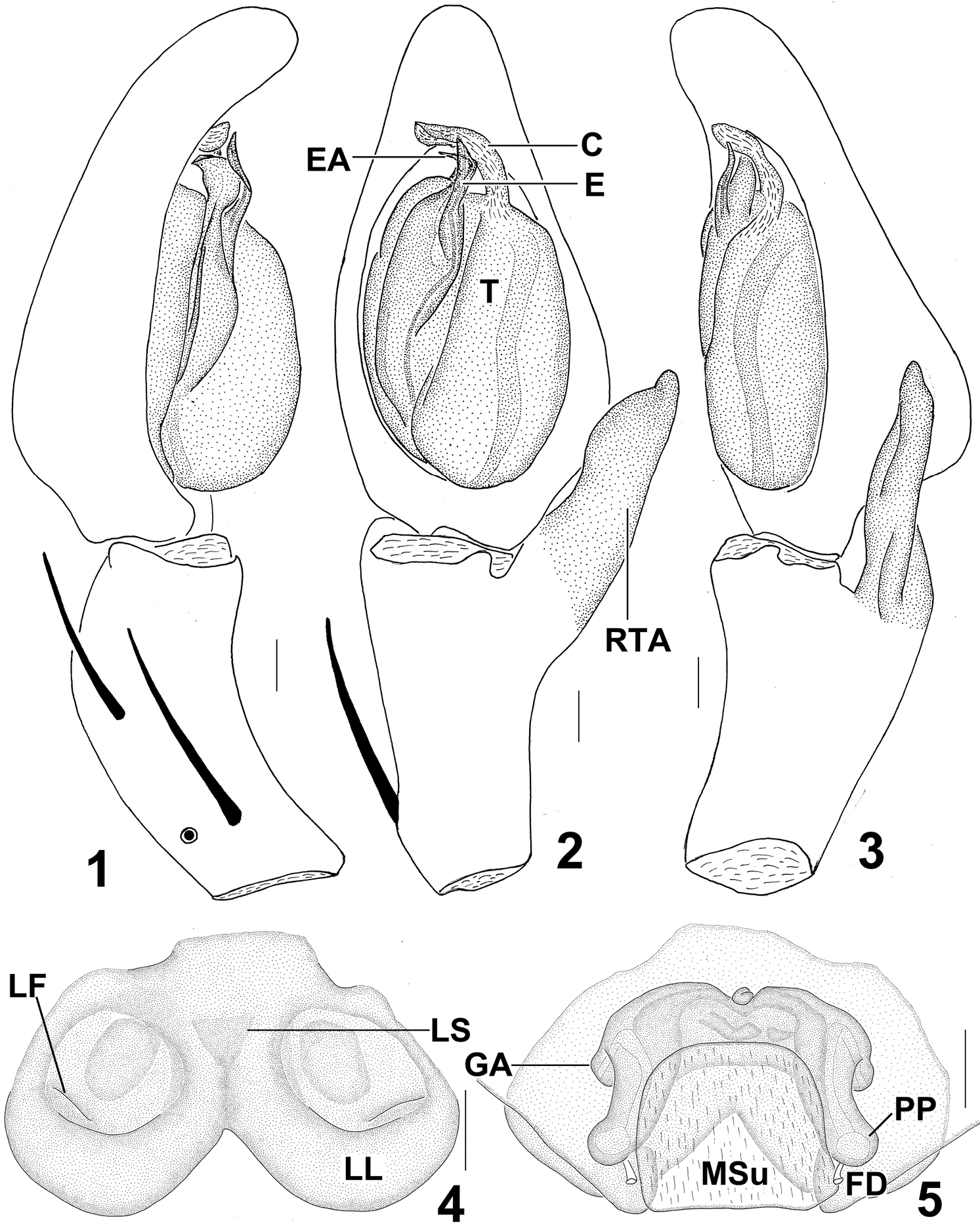
Figures 1–5.Sinopoda serrata (Wang, 1990), from Tiantangzhai National Forest Park (Hubei Province, China). 1 Left male palp, prolateral view 2 Left male palp, ventral view 3 Left male palp, retrolateral view 4 Epigyne, ventral view 5 Vulva, dorsal view. Scales = 0.2 mm. C conductor, E embolus, EA embolic apophysis, FD fertilization duct, GA glandular appendage, LF lateral furrow, LL lateral lobes, LS lobal septum, MSu membranous sac unexpanded, RTA retrolateral tibial apophysis, PP posterior part of spermathecae, T tegulum.
Included On The Following Pages:
- Life (creatures)
- Cellular (cellular organisms)
- Eukaryota (eukaryotes)
- Opisthokonta (opisthokonts)
- Metazoa (Animal)
- Bilateria
- Protostomia (protostomes)
- Ecdysozoa (ecdysozoans)
- Arthropoda (arthropods)
- Chelicerata (chelicerates)
- Arachnida (arachnids)
- Araneae (spiders)
- Opisthothelae
- Araneomorphae
- Entelegynae
- Retrolateral tibial apophysis
- Sparassidae (huntsman spiders)
- Sinopoda
- Sinopoda serrata
- Panarthropoda
This image is not featured in any collections.
Source Information
- license
- cc-by-3.0
- copyright
- Dan Quan, Jian Chen, Jie Liu
- bibliographic citation
- Quan D, Chen J, Liu J (2013) First description of the female of Sinopoda serrata (Wang, 1990) (Araneae, Sparassidae) ZooKeys 321: 89–96
- original
- original media file
- visit source
- partner site
- Zookeys
- ID