Image of Dicondylia
Description:
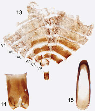
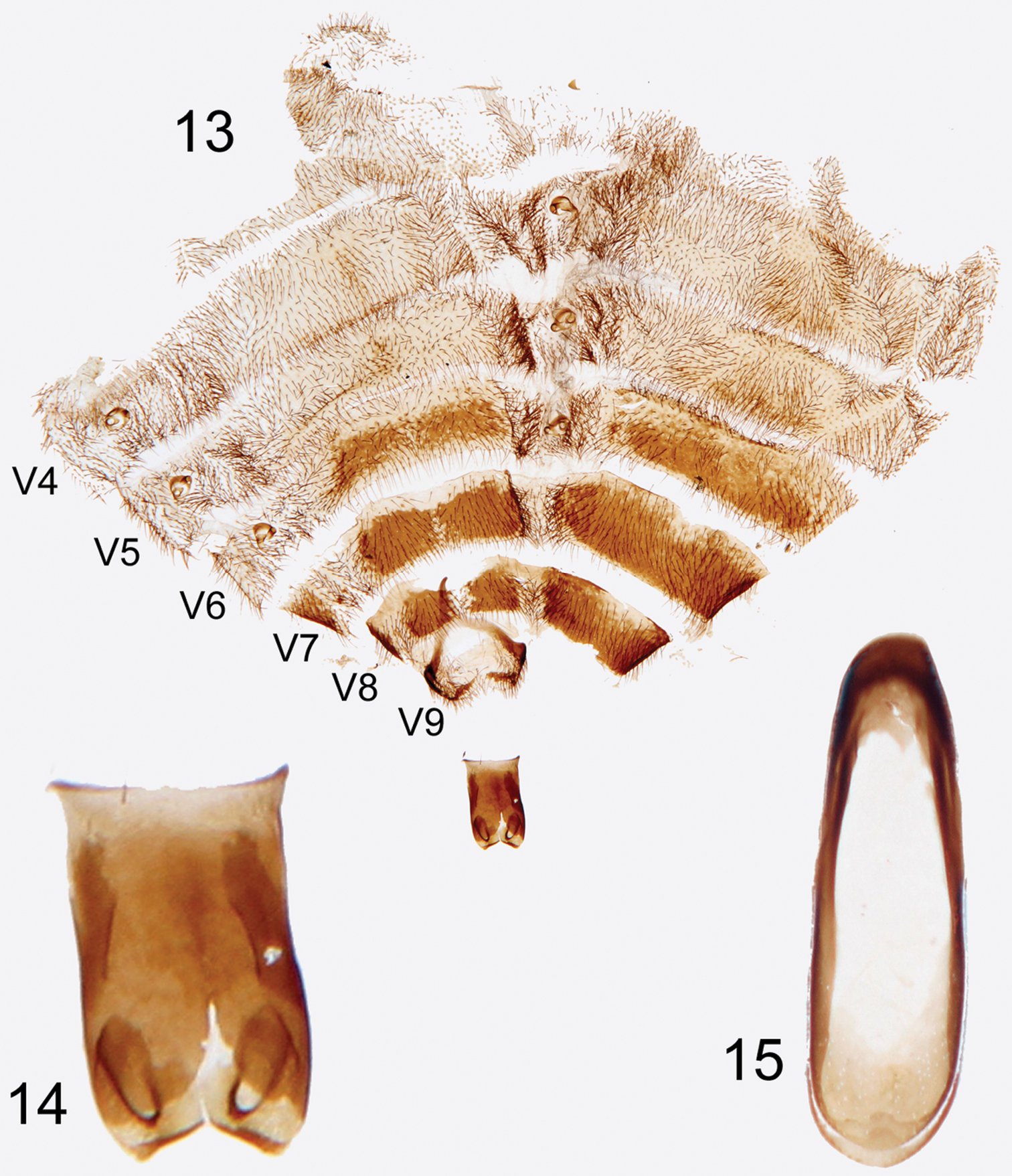
Figures 13–15.Photographs of slide mounted abdominal structures from holotype male of Rhipidocyrtus muiri Falin & Engel, gen. et sp. n. 13 Splayed abdomen as preserved on slide, numbered ventrites to the left, unnumbered tergites to the right 14 Enlarged detail, ventral view of tegmen 15 Enlarged detail of median lobe.
Included On The Following Pages:
- Biota
- Eukaryota (eukaryotes)
- Unikonta
- Opisthokonta (opisthokonts)
- Metazoa (animals)
- Epitheliozoa
- Eumetazoa
- Bilateria
- Protostomia (protostomes)
- Ecdysozoa (ecdysozoans)
- Arthropoda (arthropods)
- Hexapoda (hexapods)
- Insecta (insects)
- Dicondylia
- Pterygota (winged insects)
- Metapterygota
- Neoptera (neopteran)
- Eumetabola
- Endopterygota (endopterygotes)
- Coleoptera (beetles)
- Polyphaga
- Cucujiformia
- Tenebrionoidea (Fungus, Bark, Darkling and Blister Beetles)
- Ripiphoridae (ripiphorid beetles)
- Ripidiinae
- Ripidiini
- Rhipidocyrtus
- Rhipidocyrtus muiri
This image is not featured in any collections.
Source Information
- license
- cc-by-3.0
- copyright
- Zachary H. Falin, Michael S. Engel
- bibliographic citation
- Falin Z, Engel M (2014) Serendipity at the Smithsonian: The 107-year journey of Rhipidocyrtus muiri Falin & Engel, new genus and species (Ripidiinae, Ripidiini), from jungle beast to valid taxon ZooKeys 424: 101–116
- original
- original media file
- visit source
- partner site
- Zookeys
- ID