Bacillus subtilis image
Description:
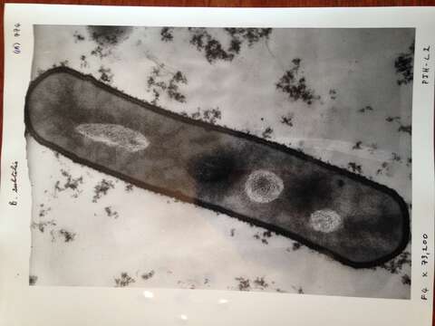
Description: English: An electron-micrograph of a section of the bacterium Bacillus subtilis (x73,200). This is a rod-shaped single cell organism, and the section is a slice (about 50nm thick) through the cell, containing the central axis of the cell. Date: 1968. Source: Estate of Peter Highton a molecular biologist working at University of Edinburgh 1968-1990. Author: Peter Highton.
Included On The Following Pages:
- Life
- Cellular
- Bacteria
- Firmicutes (gram-positive bacteria)
- Bacilli
- Bacillales
- Bacillaceae
- Bacillus
- Bacillus subtilis
- Bacillus subtilis
This image is not featured in any collections.
Source Information
- license
- cc-publicdomain
- creator
- Peter Highton
- source
- Estate of Peter Highton a molecular biologist working at University of Edinburgh 1968-1990
- original
- original media file
- visit source
- partner site
- Wikimedia Commons
- ID