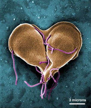
Giardia lamblia in a late stage of cell division
Description:
Description: English: This digitally-colorized scanning electron micrograph (SEM) depicted a Giardia lamblia protozoan that was about to become two, separate organisms, as it was caught in a late stage of cell division, producing a heart-shaped form. The protozoan Giardia causes the diarrheal disease called giardiasis. Giardia species exist as free-swimming (by means of flagella) trophozoites, and as egg-shaped cysts. It is the cystic stage, which facilitates the survival of these organisms under harsh environmental conditions. The cyst is considered the infective form, and disease is often transmitted by drinking contaminated water. As depicted in these SEMs, in the intestine, cysts are stimulated to liberate trophozoites. Cysts can be shed in fecal material, and can, thereafter, remain viable for several months in appropriate environmental conditions. Cysts can also be transferred directly from person-to-person, as a result of poor hygiene. Date: 1999. Source: https://phil.cdc.gov/phil/details.asp?pid=11652. Author: Dr. Stan Erlandsen.
Included On The Following Pages:
- Life (creatures)
- Cellular (cellular organisms)
- Eukaryota (eukaryotes)
- Excavates (excavates)
- Metamonada (metamonad)
- Fornicata
- Diplomonadida
- Hexamitidae
- Giardiinae
- Giardia
- Giardia intestinalis
This image is not featured in any collections.
Source Information
- license
- cc-publicdomain
- creator
- Dr. Stan Erlandsen
- source
- https://phil.cdc.gov/phil/details.asp?pid=11652
- original
- original media file
- visit source
- partner site
- Wikimedia Commons
- ID