Echinocyamus pusillus micrographs
Description:
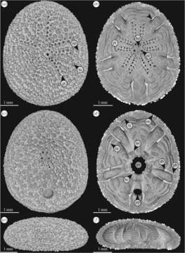
Description: English: Figure 1. Echinocyamus pusillus. Micro-CT-based volume rendering of the denuded test [GPIT/EC/00740:gg-al-1.73]. (a) View of the aboral side. (b) Horizontal section with view onto the internal aboral surface. (c) View of the oral side. (d) Horizontal section with view onto the internal oral surface. (e) Lateral view, anterior to the left. (f) Lateral section showing internal test structures. Abbreviations: au, auricle; bc, basicoronal ring; bu, buttress; db, distal buttress; gp, genital pore; gt, glassy tubercle; hp, hydropore; hr, horizontal rib; is, inner surface; os, outer surface; pb, proximal buttress; ps, peristome; pt, petal; pp, periproct; tu,tubercle; up, unipore. Date: Published 9 May 2018.DOI: 10.1098/rsos.171323. Source: http://rsos.royalsocietypublishing.org/content/5/5/171323. Author: Tobias B. Grun, James H. Nebelsick.
Included On The Following Pages:
- Life (creatures)
- Cellular (cellular organisms)
- Eukaryota (eukaryotes)
- Opisthokonta (opisthokonts)
- Metazoa (Animal)
- Bilateria
- Deuterostomia (deuterostomes)
- Echinodermata (echinoderms)
- Echinozoa
- Echinoidea (sea urchins)
- Euechinoidea
- Irregularia
- Neognathostomata
- Clypeasteroida (Sand dollars)
- Scutellina
- Laganiformes
- Fibulariidae
- Echinocyamus
- Echinocyamus pusillus (broad beau of sea)
This image is not featured in any collections.
Source Information
- license
- cc-by-sa-3.0
- copyright
- Tobias B. Grun, James H. Nebelsick
- creator
- Tobias B. Grun, James H. Nebelsick
- source
- http://rsos.royalsocietypublishing.org/content/5/5/171323
- original
- original media file
- visit source
- partner site
- Wikimedia Commons
- ID