Lax 33014 elife-33014-fig2C-v1
Description:
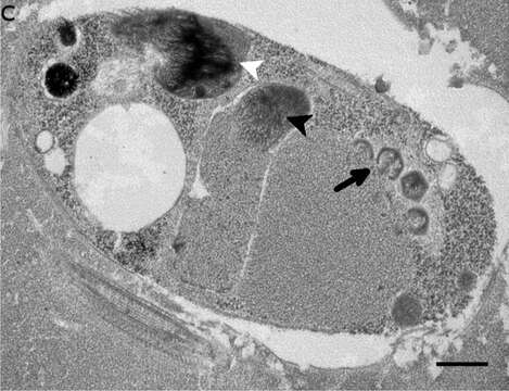
Summary.mw-parser-output table.commons-file-information-table,.mw-parser-output.fileinfotpl-type-information{border:1px solid #a2a9b1;background-color:#f8f9fa;padding:5px;font-size:95%;border-spacing:2px;box-sizing:border-box;margin:0;width:100%}.mw-parser-output table.commons-file-information-table>tbody>tr,.mw-parser-output.fileinfotpl-type-information>tbody>tr{vertical-align:top}.mw-parser-output table.commons-file-information-table>tbody>tr>td,.mw-parser-output table.commons-file-information-table>tbody>tr>th,.mw-parser-output.fileinfotpl-type-information>tbody>tr>td,.mw-parser-output.fileinfotpl-type-information>tbody>tr>th{padding:4px}.mw-parser-output.fileinfo-paramfield{background:#ccf;text-align:right;padding-right:0.4em;width:15%;font-weight:bold}.mw-parser-output.commons-file-information-table+table.commons-file-information-table,.mw-parser-output.commons-file-information-table+div.commons-file-information-table>table{border-top:0;padding-top:0;margin-top:-8px}@media only screen and (max-width:719px){.mw-parser-output table.commons-file-information-table,.mw-parser-output.commons-file-information-table.fileinfotpl-type-information{border-spacing:0;padding:0;word-break:break-word;width:100%!important}.mw-parser-output.commons-file-information-table>tbody,.mw-parser-output.fileinfotpl-type-information>tbody{display:block}.mw-parser-output.commons-file-information-table>tbody>tr>td,.mw-parser-output.commons-file-information-table>tbody>tr>th,.mw-parser-output.fileinfotpl-type-information>tbody>tr>td,.mw-parser-output.fileinfotpl-type-information>tbody>tr>th{padding:0.2em 0.4em;text-align:left;text-align:start}.mw-parser-output.commons-file-information-table>tbody>tr,.mw-parser-output.fileinfotpl-type-information>tbody>tr{display:flex;flex-direction:column}.mw-parser-output.commons-file-information-table+table.commons-file-information-table,.mw-parser-output.commons-file-information-table+div.commons-file-information-table>table{margin-top:-1px}.mw-parser-output.fileinfo-paramfield{box-sizing:border-box;flex:1 0 100%;width:100%}} Description: English: Ultrastructure of Bodo saltans virus (BsV) particles and replication. Cell of Bodo saltans 24 hr post-BsV infection: Most subcellular compartments of healthy cells have been displaced by the virus factory now taking up a third of the cell. Virion production is directed toward the periphery of the cell (black arrow). Kinetoplast genome remains intact (white arrow head) while the nuclear genome is degraded (black arrow head; Scale bar = 500 nm). Date: 27 March 2018. Source: https://elifesciences.org/articles/33014/figures at https://elifesciences.org/articles/33014 The kinetoplastid-infecting Bodo saltans virus (BsV), a window into the most abundant giant viruses in the sea. In: eLife 2018;7:e33014 doi:10.7554/eLife.33014 . Author: Christoph M. Deeg, Cheryl-Emiliane T. Chow, Curtis A. Suttle. Other versions: .
Included On The Following Pages:
- Life
- Cellular
- Eukaryota (eukaryotes)
- Excavates (excavates)
- Discoba (Jakobids)
- Euglenozoa
- Kinetoplastea
- Metakinetoplastina
- Eubodonida
- Bodonidae
- Bodo
- Bodo saltans
This image is not featured in any collections.
Source Information
- license
- cc-by-sa-3.0
- copyright
- Christoph M. Deeg, Cheryl-Emiliane T. Chow, Curtis A. Suttle
- creator
- Christoph M. Deeg, Cheryl-Emiliane T. Chow, Curtis A. Suttle
- source
- https://elifesciences.org/articles/33014/figures at https://elifesciences.org/articles/33014 The kinetoplastid-infecting Bodo saltans virus (BsV), a window into the most abundant giant viruses in the sea. In: eLife 2018;7:e33014 doi:10.7554/eLife.33014
- original
- original media file
- visit source
- partner site
- Wikimedia Commons
- ID