Bacteriophage T7 Structural Model at Atomic Resolution
Description:
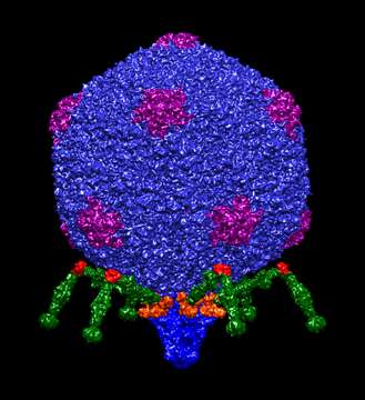
Summary.mw-parser-output table.commons-file-information-table,.mw-parser-output.fileinfotpl-type-information{border:1px solid #a2a9b1;background-color:#f8f9fa;padding:5px;font-size:95%;border-spacing:2px;box-sizing:border-box;margin:0;width:100%}.mw-parser-output table.commons-file-information-table>tbody>tr,.mw-parser-output.fileinfotpl-type-information>tbody>tr{vertical-align:top}.mw-parser-output table.commons-file-information-table>tbody>tr>td,.mw-parser-output table.commons-file-information-table>tbody>tr>th,.mw-parser-output.fileinfotpl-type-information>tbody>tr>td,.mw-parser-output.fileinfotpl-type-information>tbody>tr>th{padding:4px}.mw-parser-output.fileinfo-paramfield{background:#ccf;text-align:right;padding-right:0.4em;width:15%;font-weight:bold}.mw-parser-output.commons-file-information-table+table.commons-file-information-table,.mw-parser-output.commons-file-information-table+div.commons-file-information-table>table{border-top:0;padding-top:0;margin-top:-8px}@media only screen and (max-width:719px){.mw-parser-output table.commons-file-information-table,.mw-parser-output.commons-file-information-table.fileinfotpl-type-information{border-spacing:0;padding:0;word-break:break-word;width:100%!important}.mw-parser-output.commons-file-information-table>tbody,.mw-parser-output.fileinfotpl-type-information>tbody{display:block}.mw-parser-output.commons-file-information-table>tbody>tr>td,.mw-parser-output.commons-file-information-table>tbody>tr>th,.mw-parser-output.fileinfotpl-type-information>tbody>tr>td,.mw-parser-output.fileinfotpl-type-information>tbody>tr>th{padding:0.2em 0.4em;text-align:left;text-align:start}.mw-parser-output.commons-file-information-table>tbody>tr,.mw-parser-output.fileinfotpl-type-information>tbody>tr{display:flex;flex-direction:column}.mw-parser-output.commons-file-information-table+table.commons-file-information-table,.mw-parser-output.commons-file-information-table+div.commons-file-information-table>table{margin-top:-1px}.mw-parser-output.fileinfo-paramfield{box-sizing:border-box;flex:1 0 100%;width:100%}} Description: English: Bacteriophage t7 structural model at atomic resolution. This structural model has been constructed in UCSF Chimera software putting together all the structures that compose bacteriophage t7 using cryoEM reconstructions and pdb structures. By Dr. Victor Padilla-Sanchez, PhD from Catholic University of America. 70padillasan@cua.edu Cite as: Dr. Victor Padilla-Sanchez, PhD. (2021). Bacteriophage T7 Structural Model at Atomic Resolution. Zenodo. http://doi.org/10.5281/zenodo.5090262. Date: 10 July 2021. Source: Own work. Author: Dr. Victor Padilla-Sanchez, PhD.
Included On The Following Pages:
- Life (creatures)
- Viruses
- Caudovirales
- Autographiviridae
- Studiervirinae
- Teseptimavirus
- Escherichia virus T7
This image is not featured in any collections.
Source Information
- license
- cc-by-sa-3.0
- copyright
- Dr. Victor Padilla-Sanchez, PhD
- creator
- Dr. Victor Padilla-Sanchez, PhD
- original
- original media file
- visit source
- partner site
- Wikimedia Commons
- ID