Microsporaceae

Description:
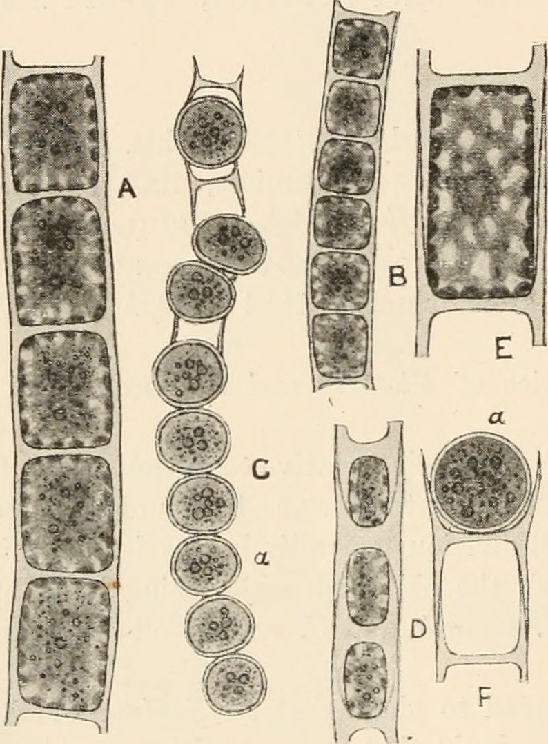
Description: English: Fig. 184. A, Microspora amœna (Kütz.) Lagerh. B and C, ? M. abbreriata (Rabenh.) Lagerh.; B, vegetative filament; C, filament with aplanospores (a). D, M. pachyderma (Wille) Lagerh. E, single vegetative cell of M. amœna var. crassior Hansg., showing the reticulated chloroplast. The indistinct blur in the centre of the cell indicates the position of the nucleus. F, fragment of filament of M. amœna with aplanospore (a). All x 520. Date: 1916. Source: https://www.flickr.com/photos/126377022@N07/14760704711/ page 301 of "Algæ. Vol. I. Myxophyceæ, Peridinieæ, Bacillarieæ, Chlorophyceæ, together with a brief summary of the occurrence and distribution of freshwat4er Algæ" (1916). Author: West, G. S. (George Stephen), 1876-1919.
Included On The Following Pages:
- Life
- Cellular
- Eukaryota
- Archaeplastida (plants)
- Chloroplastida
- Chlorophyta
- Chlorophyceae
- Microsporaceae
- Microspora
This image is not featured in any collections.
Source Information
- license
- cc-publicdomain
- creator
- West, G. S. (George Stephen), 1876-1919
- source
- Internet Archive Book Images (126377022@N07)
- original
- original media file
- visit source
- partner site
- Wikimedia Commons
- ID