323022 1 En 5 Fig3ur HTML
Description:
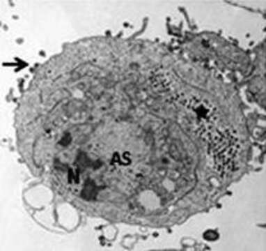
Description: English: Transmission electron micrographs of FHM cells infected by Frog virus 3 (FV3). Typical virus-infected cells with nuclei (N) showing evidence of chromatin condensation, well-defined viral assembly sites (AS); intracytoplasmic paracrystalline arrays (asterisk), and virions budding from the plasma membrane (arrow). Date: 2015. Source: Fig. 3 upper right at https://link.springer.com/chapter/10.1007/978-3-319-13755-1_5 Ranavirus Replication: Molecular, Cellular, and Immunological Events. In: Ranavirus Replication: Molecular, Cellular, and Immunological Events. In: Gray M., Chinchar V. (eds) Ranaviruses. Springer, Cham. doi:10.1007/978-3-319-13755-1_5 . Author: Jancovich J.K., Qin Q., Zhang QY., Chinchar V.G; Gray M., Chinchar V. (eds). Other versions: .
Included On The Following Pages:
- Life (creatures)
- Viruses
- Pimascovirales
- Iridoviridae (invertebrate iridescent virus and relatives)
- Alphairidovirinae
- Ranavirus
This image is not featured in any collections.
Source Information
- license
- cc-by-sa-3.0
- copyright
- Jancovich J.K., Qin Q., Zhang QY., Chinchar V.G; Gray M., Chinchar V. (eds)
- creator
- Jancovich J.K., Qin Q., Zhang QY., Chinchar V.G; Gray M., Chinchar V. (eds)
- source
- Fig. 3 upper right at https://link.springer.com/chapter/10.1007/978-3-319-13755-1_5 Ranavirus Replication: Molecular, Cellular, and Immunological Events. In: Ranavirus Replication: Molecular, Cellular, and Immunological Events. In: Gray M., Chinchar V. (eds) Ranaviruses. Springer, Cham. doi:10.1007/978-3-319-13755-1_5
- original
- original media file
- visit source
- partner site
- Wikimedia Commons
- ID