F19-01-9780123846846.jpg-550x0
Description:
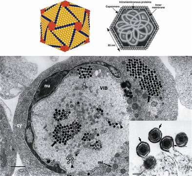
Description: English: (Top left) Outer shell of Invertebrate iridescent virus 2 (IIV-2, genus Iridovirus) (from Wrigley et al. (1969). J. Gen. Virol., 5, 123; with permission). (Top right) Schematic diagram of a cross-section of an Iridovirus particle, showing capsomers, transmembrane proteins within the lipid bilayer, and an internal filamentous nucleoprotein core (from Darcy-Tripier et al. (1984). Virology, 138, 287; with permission). (Bottom left) Transmission electron micrograph of a fat head minnow cell infected with an isolate of European catfish virus (ECV, genus Ranavirus). Nucleus (nu); virus inclusion body (VIB); paracrystalline array of non-enveloped virus particles (arrows); incomplete nucleocapsids (arrowheads); cytoplasm (cy); mitochondrion (mi); the bar represents 1 µm (from Hyatt et al. (2000). Arch. Virol., 145, 301; with permission). (insert) Transmission electron micrograph of particles of Frog virus 3 (FV-3, genus Ranavirus), budding from the plasma membrane. Arrows and arrowheads identify the viral envelope; the bar represents 200 nm. Date: 25 September 2013. Source: Fig. 1 at https://talk.ictvonline.org/ictv-reports/ictv_9th_report/dsdna-viruses-2011/w/dsdna_viruses/114/iridoviridae-figures ICTV 9th Report (2011): Iridoviridae - Figures. Author: ICTV: Jancovich, J.K., Chinchar, V.G., Hyatt, A., Miyazaki, T., Williams, T., Zhang, Q.Y. — Original authors: Wrigley, Darcy-Tripier, Hyatt et al. Other versions: .
.jpg)
.jpg)
.jpg)
Included On The Following Pages:
- Life (creatures)
- Viruses
- Pimascovirales
- Iridoviridae (invertebrate iridescent virus and relatives)
- Alphairidovirinae
- Ranavirus
This image is not featured in any collections.
Source Information
- license
- cc-by-sa-3.0
- copyright
- ICTV: Jancovich, J.K., Chinchar, V.G., Hyatt, A., Miyazaki, T., Williams, T., Zhang, Q.Y. — Original authors: Wrigley, Darcy-Tripier, Hyatt et al.
- creator
- ICTV: Jancovich, J.K., Chinchar, V.G., Hyatt, A., Miyazaki, T., Williams, T., Zhang, Q.Y. — Original authors: Wrigley, Darcy-Tripier, Hyatt et al.
- source
- Fig. 1 at https://talk.ictvonline.org/ictv-reports/ictv_9th_report/dsdna-viruses-2011/w/dsdna_viruses/114/iridoviridae-figures ICTV 9th Report (2011): Iridoviridae - Figures
- original
- original media file
- visit source
- partner site
- Wikimedia Commons
- ID