Chromoblastomycosis 2
Description:
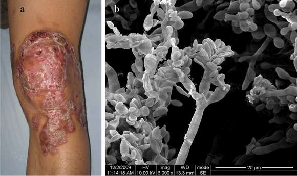
Description: a. A 34-year-old male with a 12-year history of a red plaque in the left knee. b. Under SEM observation: dematiaceous hyphae with many well-defined septa, conidiophores, and oval brown spores arranged in a clump could be seen. The surfaces of conidiogenous cells were smooth. Oval spores were arranged around conidiophores. Date: 2016. Source: [1]. Author: Ran Yuping et al.
Included On The Following Pages:
- Life
- Cellular
- Eukaryota
- Opisthokonta
- Nucletmycea
- Fungi
- Dikarya
- Ascomycota
- Eurotiomycetes
- Chaetothyriales
- Herpotrichiellaceae
- Fonsecaea
- Fonsecaea pedrosoi
This image is not featured in any collections.
Source Information
- license
- cc-by-sa-3.0
- copyright
- Ran Yuping et al.
- creator
- Ran Yuping et al.
- source
- [1]
- original
- original media file
- visit source
- partner site
- Wikimedia Commons
- ID