Mouse cingulate cortex neurons
Description:
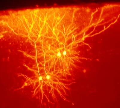
Description: English: Tyramide filled neurons from the cingulate cortex of mouse brain. The two larger cells on the left are in upper layer 5, and the two on the right are in layer 2/3. Image taken by holding a digital camera against the eyepiece of a dissecting scope. Date: 1 June 2005, 19:40. Source: tyramideFills. Author: Mark Miller from San Francisco, CA, USA.
Included On The Following Pages:
- Life (creatures)
- Cellular (cellular organisms)
- Eukaryota (eukaryotes)
- Opisthokonta (opisthokonts)
- Metazoa (Animal)
- Bilateria
- Deuterostomia (deuterostomes)
- Chordata (Chordates)
- Vertebrata (vertebrates)
- Gnathostomata (jawed fish)
- Osteichthyes
- Sarcopterygii (Lobe-finned fishes)
- Tetrapoda (terrestrial vertebrates)
- Amniota (amniotes)
- Synapsida (synapsids)
- Therapsida (therapsid)
- Cynodontia (cynodonts)
- Mammalia (mammals)
- Theria (Therians)
- Eutheria (eutherian)
- Placentalia (placental)
- Boreoeutheria
- Euarchontoglires
- Glires
- Rodentia (rodents)
- Mouse relatives
- Myomorpha (mice, rats, gerbils, jerboas, and relatives)
- Muroidea (mice, rats, gerbils, and relatives)
- Eumuroidea
- Muridae (murid rodents)
- Murinae (Murine)
- Mus (Old World Mice and Pygmy Mice)
- Mus
- Mus musculus (house mouse)
This image is not featured in any collections.
Source Information
- license
- cc-by-sa-3.0
- copyright
- Mark Miller
- creator
- Mark Miller
- source
- Flickr user ID neurollero
- original
- original media file
- visit source
- partner site
- Wikimedia Commons
- ID