Spicules and skeleton

Description:
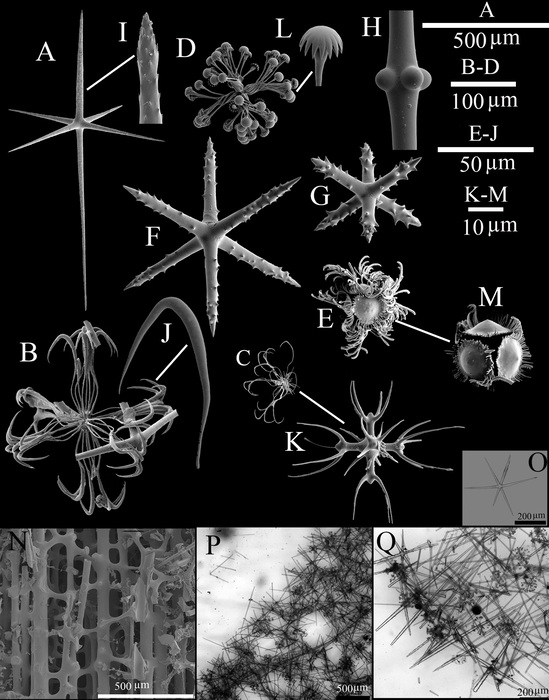
A, dermal or atrial hexactin; B, drepanocome I; C, drepanocome II; D, spirodiscohexaster; E, plumicome; F, microhexactin I; G, microhexactin II; H, detail of tubercles in the middle of a diactine; I, detail of the pinular ray of a hexactin; J, detail of the hook-like secondary ray of a drepanocome I; K, detail of the middle part of a drepanocome II; L, detail of the tooth disc of a spirodiscohexaster; M, detail of the whorl of a plumicome; N, spicules of the peduncle; O–Q, LM images of spicule and skeleton; O, choanosomal hexactin; P, tangential view of choanosomal structure; Q, transversal view of choanosomal structure.
Included On The Following Pages:
- Life
- Cellular
- Eukaryota (eukaryotes)
- Opisthokonta (opisthokonts)
- Metazoa (animals)
- Porifera (sponges)
- Hexactinellida (hexactinellid sponges)
- Hexasterophora
- Lyssacinosida
- Euplectellidae
- Bolosominae
- Saccocalyx
- Saccocalyx microhexactin
This image is not featured in any collections.
Source Information
- license
- cc-by-nc-sa-4.0
- copyright
- WoRMS Editorial Board
- contributor
- Cárdenas, Paco, P.
- original
- original media file
- visit source
- partner site
- World Register of Marine Species
- ID