Portrait
Description:
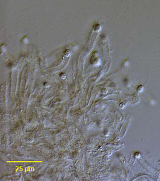
Portrait of a group of Siphomonas fritschii (Pringsheim,1946), colorless chrysophytes, Cells occur singly or in pairs at the ends of hollow, sometimes forked tubes with their flagella protruding. These tubes occur in tangles and are sometimes tinged various shades of brown due to manganese and iron salts. The cells are rounded posteriorly and slightly truncate anteriorly. Two unequal flagella arise from the edge of the anterior end. A small greenish stigma lies near the flagellar insertions. A contractile vacuole is present. The cells frequently flee their stalks to swim free. Some empty tubes are seen here. Collected from an ephemeral freshwater pool near Boise, Idaho November 2004. Previously reported only from England and France.DIC.
Included On The Following Pages:
- Life (creatures)
- Cellular (cellular organisms)
- Eukaryota (eukaryotes)
- SAR (Stramenopiles, Alveolates, Rhizaria)
- Stramenopiles (heterokont)
- Ochrophyta (Ochrophyte)
- Chrysophyceae (golden algae)
- Chromulinales
- Chromulinaceae
- Siphomonas
- Siphomonas fritschii
This image is not featured in any collections.
Source Information
- license
- cc-by-nc
- author
- William Bourland
- provider
- micro*scope
- original
- original media file
- visit source
- partner site
- micro*scope
- ID