in vivo
Description:
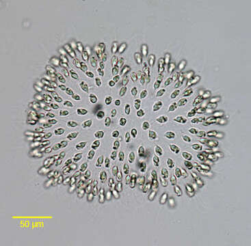
Portrait of the colonial chrysophyte, Uroglena volvox (Ehrenberg, 1835). colonies free-swimming, spherical, ellipsoidal or oblong, up to several 100 (rarely >1000)μm in diameter; cells radially arranged in a single layer at the periphery of a gelatinous matrix;the friable colonies are easily disrupted by the pressure of the coverslip. The interior of the matrix is fairly homogeneous or containing a system of fine, radiating and branched stalks to which the cells are attached by their pointed posterior ends; individual cells Ochromonas-like, with 2 flagella of unequal length; chloroplasts 1-2, laminate to discoid, at least in one species containing a pyrenoid; eyespot usally 1 (rarely 2 or lacking); contractile vacuoles 1-3; numerous muciferous bodies located at the cell periphery; nutrition phototrophic and phagotrophic; cell division longitudinal; colony reproduction by constriction into two daughter colonies or by fragmentation; stomatocysts frequently observed, their ornamentation used for species identification; some species of common occurrence in the plankton of lakes and ponds, sometimes bloom-forming, one marine species. Collected from a freshwater dredge pond near Idaho City, Idaho June 2005. Brightfield.
Included On The Following Pages:
- Life (creatures)
- Cellular (cellular organisms)
- Eukaryota (eukaryotes)
- SAR (Stramenopiles, Alveolates, Rhizaria)
- Stramenopiles (heterokont)
- Ochrophyta (Ochrophyte)
- Chrysophyceae (golden algae)
- Chromulinales
- Chromulinaceae
- Uroglena
- Uroglena volvox
This image is not featured in any collections.
Source Information
- license
- cc-by-nc
- author
- William Bourland
- provider
- micro*scope
- original
- original media file
- visit source
- partner site
- micro*scope
- ID