portrait
Description:
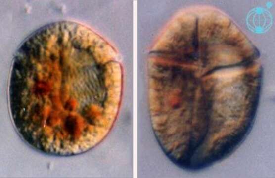
Amphidinium (am-fee-din-ee-um) corpulentum Kofoid & Swezy 1921. The image on the left is a mid focus plane through a cell showing the nucleus on the right side of the cell (= the right image side). Red to orange food vacuoles and yellow-brown plastids are visible. The image on the right shows a cell in ventral view (side-reversed). The cingulum is at the anterior end and an apical groove is visible.
Included On The Following Pages:
- Life (creatures)
- Cellular (cellular organisms)
- Eukaryota (eukaryotes)
- SAR (Stramenopiles, Alveolates, Rhizaria)
- Alveolata (alveolates)
- Dinophyceae
- Gymnodiniales
- Gymnodiniaceae
- Amphidinium
- Amphidinium corpulentum
This image is not featured in any collections.
Source Information
- license
- cc-by-nc
- author
- Mona Hoppenrath and Shauna Murray
- provider
- micro*scope
- original
- original media file
- visit source
- partner site
- micro*scope
- ID