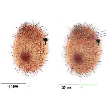
Ventrolateral view of infraciliature
Description:
Ventrolateral view of the infraciliature of the hymenostome ciliate, Dexiotricha granulosa (Kent, 1881) Foissner, 1994. Synonym of Loxocephulus granulosa. The cell is ovoid, broadly rounded posteriorly and truncate anteriorly. Regular longitudinal kineties terminate at a subapical band of circumferential kineties demarcating a cilia-free truncate apical area or frontal plate. Fibrils radiate anteriorly from the kinetids of the anteriormost paratene. There is a single long caudal cilium (green line). The oral aperture is small and difficult to visualize in vivo. It is located in the anterior quarter with a paraoral membrane on the right (red line) and 3 adoral membranelles (dark blue lines). Closely spaced basal bodies of the somatic kinaty to the right of the oral aperture form a "pseudomembrane" (light blue line). The macronucleus is spheroid and located in the mid-cell. Single contractile vacuole. From freshwater pond near Boise, Idaho. Silver carbonate stain (see Foissner, W. Europ. J. Protistol., 27:313-330;1991).Brightfield.
Included On The Following Pages:
- Life
- Cellular
- Eukaryota (eukaryotes)
- SAR (Stramenopiles, Alveolates, Rhizaria)
- Alveolata (alveolates)
- Ciliophora (ciliates)
- Intramacronucleata
- Oligohymenophorea
- Scuticociliatia
- Philasterida
- Loxocephalidae
- Dexiotricha
- Dexiotricha granulosa
This image is not featured in any collections.
Source Information
- license
- cc-by-nc
- author
- William Bourland
- provider
- micro*scope
- original
- original media file
- visit source
- partner site
- micro*scope
- ID