in vivo
Description:
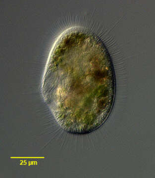
Portraitof the hymenostome ciliate, Histiobalantium natans (Claparede & Lachmann, 1858). The cell is ovoid to reniform in outline with a broadly rounded posterior. The oval oral aperture is located in the mid-portion of the cell. There is an undulating membrane on the right margin of the peristome curving around the posteriorly located cytostome to form a cup or pouch. There are three adoral membranelles on the left side of the peristome. M1 and M2 are parallel and oriented obliquely to the undulating membrane while membranelle M3 is inclined slightly posteriorly between the posterior ends of M1 and M2 and the undulating membrane, forming a triangle. The somatic ciliature is composed of longitudinal kineties. Longer bristle-like cilia are interspersed with more numerous short cilia. Both pre- and post-oral sutures are present. A long caudal cilium absent. There are three contractile vacuoles. Macronucleus with an irregular shape, usually divided into two parts with several micronuclei. Similar in appearance to Pleuronema which may have long caudal cilia but whose other somatic cilia are of equal length. Stained by the silver carbonate technique (see Foissner, W. Europ. J. Protistol., 27:313-330;1991). Collected from a freshwater pond near Boise, Idaho, february 2005.DIC.
Included On The Following Pages:
- Life
- Cellular
- Eukaryota (eukaryotes)
- SAR (Stramenopiles, Alveolates, Rhizaria)
- Alveolata (alveolates)
- Ciliophora (ciliates)
- Intramacronucleata
- Oligohymenophorea
- Scuticociliatia
- Pleuronematida
- Histiobalantiidae
- Histiobalantium
- Histiobalantium natans
This image is not featured in any collections.
Source Information
- license
- cc-by-nc
- author
- William Bourland
- provider
- micro*scope
- original
- original media file
- visit source
- partner site
- micro*scope
- ID