portrait
Description:
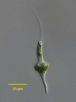
Portrait of the euglenid flagellate, Eutreptia viridis (Perty, 1852). The cells are fusiform during swimming. The pellicle shows fine spiral striations. There are two equal length emergent flagella. The cells swim rapidly with the anterior end tracing a wide circle. When cells stop swimming they exhibit marked metaboly (euglenoid movement). There are numerous bright green discoid to ellipsoid chloroplasts. Paramylon granules are rod or disc-shaped. There is a prominent red eyespot associated with one of the flagella. There is an anterior subapical opening into the reservoir. A single contractile vacuole empties into the reservoir. The nucleus is central in swimming cells. Eutreptia has been most often reported from marine and brackish habitats but is also found uncommonly in fresh water.Collected from surface samples of a slow flowing organically enriched freshwater stream overgrown with duckweed (Lemnaceae) near Boise, Idaho. DIC.
Included On The Following Pages:
- Life
- Cellular
- Eukaryota
- Excavates
- Discoba (Jakobids)
- Euglenozoa
- Euglenida (euglenoides)
- Spirocuta
- Euglenophyceae
- Eutreptiales
- Eutreptia
- Eutreptia viridis
This image is not featured in any collections.
Source Information
- license
- cc-by-nc
- author
- William Bourland
- provider
- micro*scope
- original
- original media file
- visit source
- partner site
- micro*scope
- ID