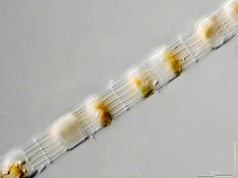
Image of Skeletonema costatum
Description:
Sampling date 01/2022. Scale bars indicate 25 µm (1, 2), 10 µm (3-6).
Three pairs of girdleband view/optical cross section in two magnification steps.
First couple:Girdleband view showing silicious connection processes, cross section showing chloroplasts and some nuclei.Second couple:Close-up.a) Girdleband view: Some ultrathin floating processes made of chitin become visible.
b) Cross section: Chloroplasts and some nuclei become clearly visabe.Third couple:Close-up.
Girdleband view: The ultrathin floating processes made of chitin are clearly visible.
These chitinous processes are characteristic of the Thalassiosirales.
Please click on < or > on the image edges or on the dots at the bottom edge of the images to browse through the slides!
Place name: Baltic Sea, Kieler Förde, Kiel Fjord (Germany)
Latitude: 54.3894126 Longitude: 10.1749055
Microscope Zeiss Axioplan, camera Olympus OM-D M5 MKII. DOF images.
© Wolfgang Bettighofer,
images under Creative Commons License V 3.0 (CC BY-NC-SA).
For permission to use of (high resolution) images please contact postmaster@protisten.de.
For further information about the image, please click here: Link to protisten.de page
Included On The Following Pages:
- Life
- Cellular
- Eukaryota (eukaryotes)
- SAR (Stramenopiles, Alveolates, Rhizaria)
- Stramenopiles (heterokont)
- Ochrophyta (Ochrophyte)
- Bacillariophyta (diatoms)
- Coscinodiscophyceae
- Thalassiosirophycidae
- Thalassiosirales
- Skeletonemataceae
- Skeletonema
- Skeletonema costatum
This image is not featured in any collections.
Source Information
- license
- cc-by-nc-sa-3.0
- copyright
- Wolfgang Bettighofer
- creator
- Wolfgang Bettighofer [email]
- original
- original media file
- visit source
- partner site
- protisten.de
- ID