Image of human body louse
Description:
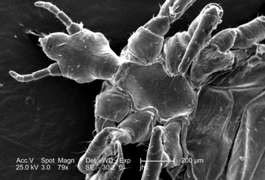
Under a relatively low magnification of 79x, this 2006 scanning electron micrograph (SEM) depicted a ventral, or inferior view, of a male louse, Pediculus humanus var. corporis. The head, or cephalic region is at the left, from which its two antennae were extended. The head attached to the thoracic region, which gave rise to its three pairs of jointed legs. While the abdominal region, towards the far right, is the region in which was housed the stomach, and intestines. The jointed nature of its extremities, designates this organism as a member of the phylum Arthropoda, and the fact that there are three pairs of legs, the louse is, thereby, placed into the class, Insecta. Note the small, hair-like structures adorning the exoskeletal surface of this insect. These are known as setae, and are not hairs at all, but extensions of the chitinous exoskeletal surface, which provide the organism with sensorial data about its surroundings.
Created: 2006
Included On The Following Pages:
- Life (creatures)
- Cellular (cellular organisms)
- Eukaryota (eukaryotes)
- Opisthokonta (opisthokonts)
- Metazoa (Animal)
- Bilateria
- Protostomia (protostomes)
- Ecdysozoa (ecdysozoans)
- Arthropoda (arthropods)
- Pancrustacea
- Hexapoda (hexapods)
- Insecta (insects)
- Pterygota (winged insects)
- Neoptera (neopteran)
- Paraneoptera
- Psocodea (bark lice, book lice and true lice)
- Troctomorpha (book louse)
- Pediculidae (primate body lice)
- Pediculus
- Pediculus humanus (human body louse)
- Panarthropoda
- Nanopsocetae
This image is not featured in any collections.
Source Information
- license
- cc-publicdomain
- photographer
- Janice Carr
- provider
- Public Health Image Library
- original
- original media file
- visit source
- partner site
- Public Health Image Library
- ID