Plate 6
Description:
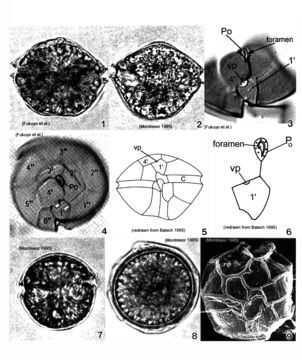
Plate 6. Alexandrium pseudogonyaulax. Figs. 1-4. LM. Fig. 1. Ventral view. Cell broadly pentagonal; wider than long. Epitheca short and dome-shaped. Hypotheca longer than epitheca. Cingulum shallow and barely displaced. Fig. 2. Dorsal view. Antapex obliquely concave. Fig. 3. Epitheca: ventral view. Apical pore plate (Po) with comma-shaped foramen. 1' plate pentagonal with large wide ventral pore (vp) on 4' plate margin. Fig. 4. Epitheca: apical view. 1' plate does not come in contact with Po. Po oval and longitudinal on apex. Figs. 5-6. Line drawings. Fig. 6. Po and 1' plate not in contact. Fig. 7. LM: isogamous gametes smaller and rounder than vegetative cells. Fig. 8. LM: round resting cyst. Fig. 9. SEM: paratabulate cyst.
Included On The Following Pages:
- Life (creatures)
- Cellular (cellular organisms)
- Eukaryota (eukaryotes)
- SAR (Stramenopiles, Alveolates, Rhizaria)
- Alveolata (alveolates)
- Dinophyceae
- Gonyaulacales
- Gonyaulacaceae
- Alexandrium
- Alexandrium pseudogonyaulax
- Dinoflagellata (dinoflagellates)
This image is not featured in any collections.
Source Information
- license
- cc-publicdomain
- bibliographic citation
- Faust, Maria A. and Rose A. Gulledge. Identifying Harmful Marine Dinoflagellates. Smithsonian Contributions from the United States National Herbarium, volume 42: 1-144 (including 48 plates, 1 figure and 1 table).
- original
- original media file
- visit source
- partner site
- NMNH Marine Dinoflagellates
- ID