NMNH Gambierdiscus belizeanus type specimen
Description:
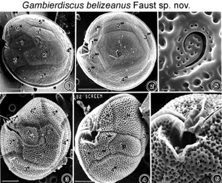
Figs. 1-6. Scanning electron micrographs of the surface morphology of Gambierdiscus toxicus and Gambierdiscus belizeanus are shown. FIGS.1-2. Scanning electron micrographs of the surface morphology of Gambierdiscus toxicus Adachi et Fukuyo. FIG.1. Cell in epithecal view. FIG.2. Cell in hypothecal view. Cell shape is round, compressed and ellipsoidal. Cell surface is smooth with scattered small pores. Thecal plate is large and quadrangular. FIGS.3-6. Gambierdiscus belizeanus sp. nov. FIG.3. Cell in epithecal view slightly damaged. Cell surface areolated and plates partially separated. FIG. 4. Cell is in hypothecal and ventral view. Plate is narrow. FIG.5. Apical pore complex is triangular with a fish-hook-shaped apical pore. A round pore is present in the areolae (arrowhead). FIG.6. Cingulum deep, ascending into a deep sulcal hollow. EMu: Holotype SEM NEGATIVE # 132003B; SEM STUB # 152; FIELD # 682-93; ACCESSION # 407167; CATALOG # 798; FIGURE # 3.
Included On The Following Pages:
- Life
- Cellular
- Eukaryota (eukaryotes)
- SAR (Stramenopiles, Alveolates, Rhizaria)
- Alveolata (alveolates)
- Dinophyceae
- Gonyaulacales
- Goniodomataceae
- Gambierdiscus
- Gambierdiscus belizeanus
- Dinoflagellata (dinoflagellates)
This image is not featured in any collections.
Source Information
- license
- cc-by-nc-sa-3.0
- copyright
- National Museum of Natural History, Smithsonian Institution
- original
- original media file
- visit source
- partner site
- NMNH Marine Dinoflagellates
- ID