Plate 29
Description:
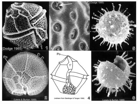
Plate 29. Lingulodinium polyedrum. Figs. 1-3. SEM. Fig. 1. Ventral view: cells angular and polyhedral-shaped. Thick plates well defined and coarsely areolate. Epitheca with shoulders and nearly flattened apex. Hypotheca with straight sides and flattened antapex (arrow). Cingulum deep and displaced 1-2 X its width. Sulcus widens posteriorly. Fig. 2. Apical view: first apical plate (1') long and narrow. Apical pore plate (Po) with raised inner elliptical ridge. Cingulum with lists (arrowheads). Strong ridges along sutures outline thecal plates. Fig. 3. Thecal areolae with large trichocysts (arrow)(Lewis and Burton 1988). Fig. 4. Line drawing. Figs. 5-6. SEM: resting cysts. Fig. 5. Cyst sperical with numerous tapering spines. Fig. 6. Cyst theca after excystment.
Included On The Following Pages:
- Life
- Cellular
- Eukaryota
- SAR (Stramenopiles, Alveolates, Rhizaria)
- Alveolata
- Dinophyceae
- Gonyaulacales
- Lingulodinium
- Lingulodinium polyedrum
- Gonyaulacaceae
- Dinoflagellata
This image is not featured in any collections.
Source Information
- license
- cc-publicdomain
- bibliographic citation
- Faust, Maria A. and Rose A. Gulledge. Identifying Harmful Marine Dinoflagellates. Smithsonian Contributions from the United States National Herbarium, volume 42: 1-144 (including 48 plates, 1 figure and 1 table).
- original
- original media file
- visit source
- partner site
- NMNH Marine Dinoflagellates
- ID