NMNH Ostreopsis belizeanus type specimen
Description:
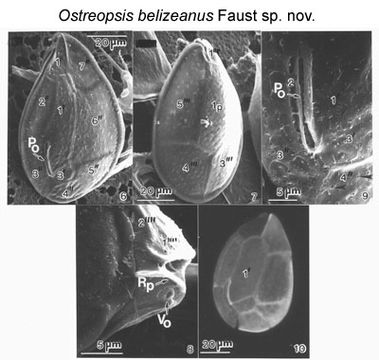
Figs 6-10. Cells of Ostreopsis belizeanus sp. nov. Figs 6-9. Scanning electron microscopy. Fig. 6. Morphology of epithecal plates and position of apical pore plate (Po). Fig. 7. Hypothecal plates. Fig. 8. In the cingulum, the ventral opening (Vo) is located adjacent to a ridged plate (Rp). Fig. 9. Apical pore plate includes a narrow apical pore (Po) located off-center. Thecal surface laced with round pores (arrows). Fig. 10. Epifluorescence light microscopy of epithecal plates. EMu: HOLOTYPE SEM NEGATIVE # 211053; SEM STUB # 211; FIELD # 1005-96; ACCESSION # 2002408; CATALOG # 1541; FIGURE # 6.
Included On The Following Pages:
This image is not featured in any collections.
Source Information
- license
- cc-by-nc-sa-3.0
- copyright
- National Museum of Natural History, Smithsonian Institution
- original
- original media file
- visit source
- partner site
- NMNH Marine Dinoflagellates
- ID