Plate 32
Description:
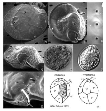
Plate 32. Ostreopsis lenticularis. Figs. 1-5. SEM. Fig. 1. Epithecal view: cell lenticulate to broadly oval. Curved off-center apical pore plate with a slit-like apical pore (arrow). Plate 1' irregularly pentagonal. Fig. 2. Hypothecal view: plate 1p central and pentagonal. Fig. 3. Smooth thecal surface. Round pores with smooth raised edges. Fig. 4. Hypothecal ventral view: cell anterio-posteriorly compressed. Shallow cingulum with smooth edge. Small sulcus hidden (arrow). Fig. 5. Location of ventral opening (arrow), ventral plate (asterisk), and rigid plate (arrowheads) within cingulum. Fig. 6. Line drawing: thecal plate arrangement. Figs. 7,8. LM. Fig. 7. Cytoplasma granulated; posterior nucleus (n). Fig. 8. Distinct cingular list.
Included On The Following Pages:
- Life
- Cellular
- Eukaryota
- SAR (Stramenopiles, Alveolates, Rhizaria)
- Alveolata
- Dinophyceae
- Ostreopsidaceae
- Ostreopsis
- Ostreopsis lenticularis
- Gonyaulacales
- Dinoflagellata
This image is not featured in any collections.
Source Information
- license
- cc-publicdomain
- bibliographic citation
- Faust, Maria A. and Rose A. Gulledge. Identifying Harmful Marine Dinoflagellates. Smithsonian Contributions from the United States National Herbarium, volume 42: 1-144 (including 48 plates, 1 figure and 1 table).
- original
- original media file
- visit source
- partner site
- NMNH Marine Dinoflagellates
- ID