Plate 34
Description:
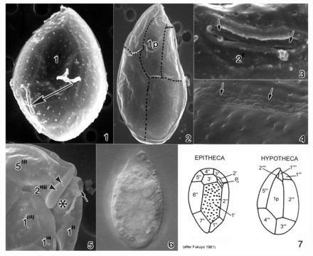
Plate 34. Ostreopsis ovata. Figs. 1-5. SEM. Fig. 1. Epithecal view: cell slender and tear-shaped. Apical pore plate (Po) off-center (arrow). Plate 1' large and hexagonal. Cingulum wide with narrow lists. Fig. 2. Hypothecal view: plates delicate. Plate 1p long and narrow. Fig. 3. Po: short and straight, adjacent to plate 2'. Fig. 4. Thecal surface smooth with scattered small pores. Suture line uneven and bumpy (arrows). Fig. 5. Hypothecal view: ventral opening (arrow), ventral plate (asterisk), and rigid plate (arrowhead) on cingulum. Fig. 6. LM. Large posterior nucleus. Fig. 7. Line drawing: thecal plate arrangement.
Included On The Following Pages:
- Life
- Cellular
- Eukaryota
- SAR (Stramenopiles, Alveolates, Rhizaria)
- Alveolata
- Dinophyceae
- Ostreopsidaceae
- Ostreopsis
- Ostreopsis ovata
- Gonyaulacales
- Dinoflagellata
This image is not featured in any collections.
Source Information
- license
- cc-publicdomain
- bibliographic citation
- Faust, Maria A. and Rose A. Gulledge. Identifying Harmful Marine Dinoflagellates. Smithsonian Contributions from the United States National Herbarium, volume 42: 1-144 (including 48 plates, 1 figure and 1 table).
- original
- original media file
- visit source
- partner site
- NMNH Marine Dinoflagellates
- ID