-
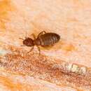
Figure 1.Photograph of habitus of Libanopsyllipsocus alexanderasnitsyni gen. et sp. n., holotype, male, specimen number 30.
-
Saima Naz, Oldrich Sychra, Syed Anser Rizvi
Zookeys
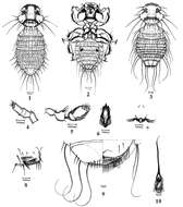
Figures 1–10.Colpocephalum afrozeae sp. n.1 male dorsal view 2 male ventral view 3 female dorsal view 4 maxillary palp 5 antenna; 6 hypopharynx 7 prosternal plate 8 sternite IV with ctenidia 9 female terminalia ventral view 10 male genitalia
-
Figure 10.Drawing of hypandrium of Libanopsyllipsocus alexanderasnitsyni gen. et sp. n., holotype, male; par = paraproct, ep = epiproct, trich = trichobothria; scale bar = 0.3 mm.
-
Saima Naz, Oldrich Sychra, Syed Anser Rizvi
Zookeys
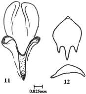
Figures 11–12.Colpocephalum afrozeae sp. n. 11 penis details 12 genital sclerite.
-
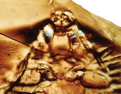
Figure 11.Photograph of hypandrium and aedeagus of Libanopsyllipsocus alexanderasnitsyni gen. et sp. n., holotype, male.
-
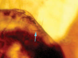
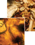
Figure 12.Photograph of paraproct of Libanopsyllipsocus alexanderasnitsyni gen. et sp. n., holotype, male; arrow shows the anal spine.
-
Figure 2.Drawing of habitus of Libanopsyllipsocus alexanderasnitsyni gen. et sp. n., holotype, male, scale bar = 0.3 mm.
-
Figure 3.Photograph of hypopharynx filaments (arrows) of Libanopsyllipsocus alexanderasnitsyni gen. et sp. n., holotype, male.
-
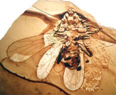
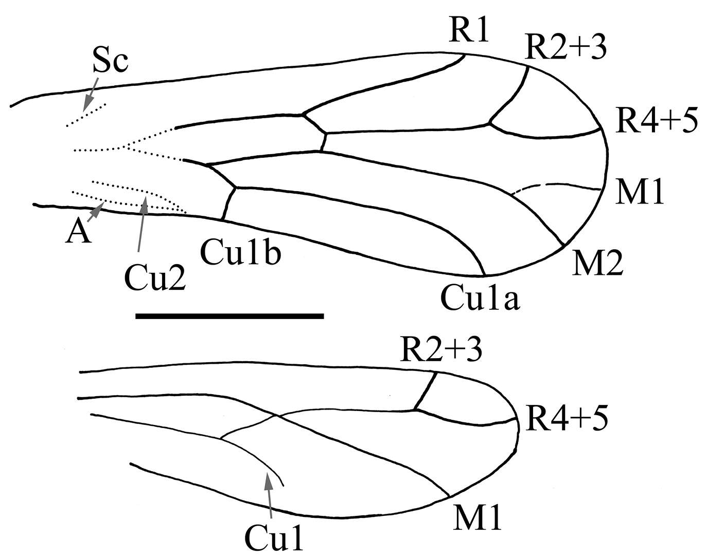
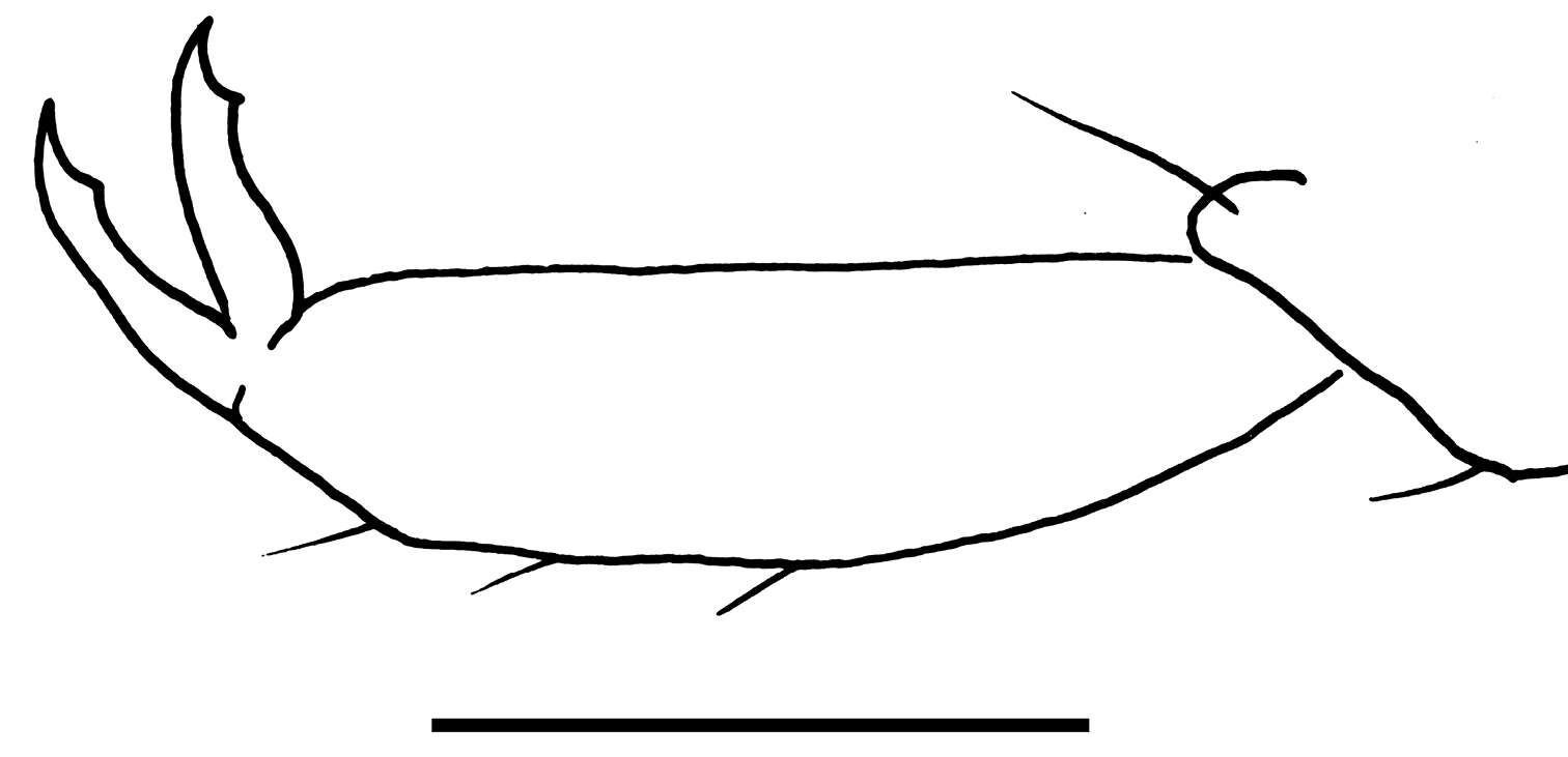
Figure 4.Drawing of wings of Libanopsyllipsocus alexanderasnitsyni gen. et sp. n., holotype, male, scale bar = 0.3 mm.
-
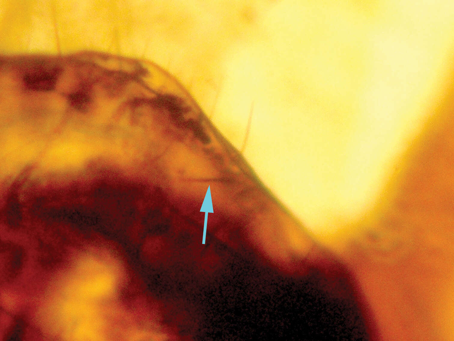
Figure 5.Microphotograph of nodulus, arrow showing the meeting area of Cu2 and A.
-
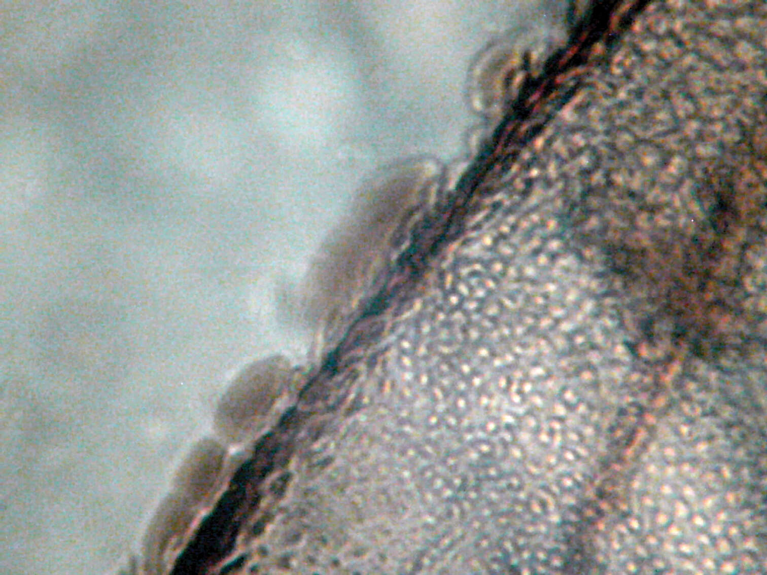
Figure 6.Microphotograph of structure of forewing margin.
-
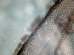
Figure 7.Microphotograph of hind leg coxal rasp (Pearman’s organ).
-
Figure 8.Drawing of pretarsal claw of Libanopsyllipsocus alexanderasnitsyni gen. et sp. n., holotype, male, scale bar = 0.03 mm.
-
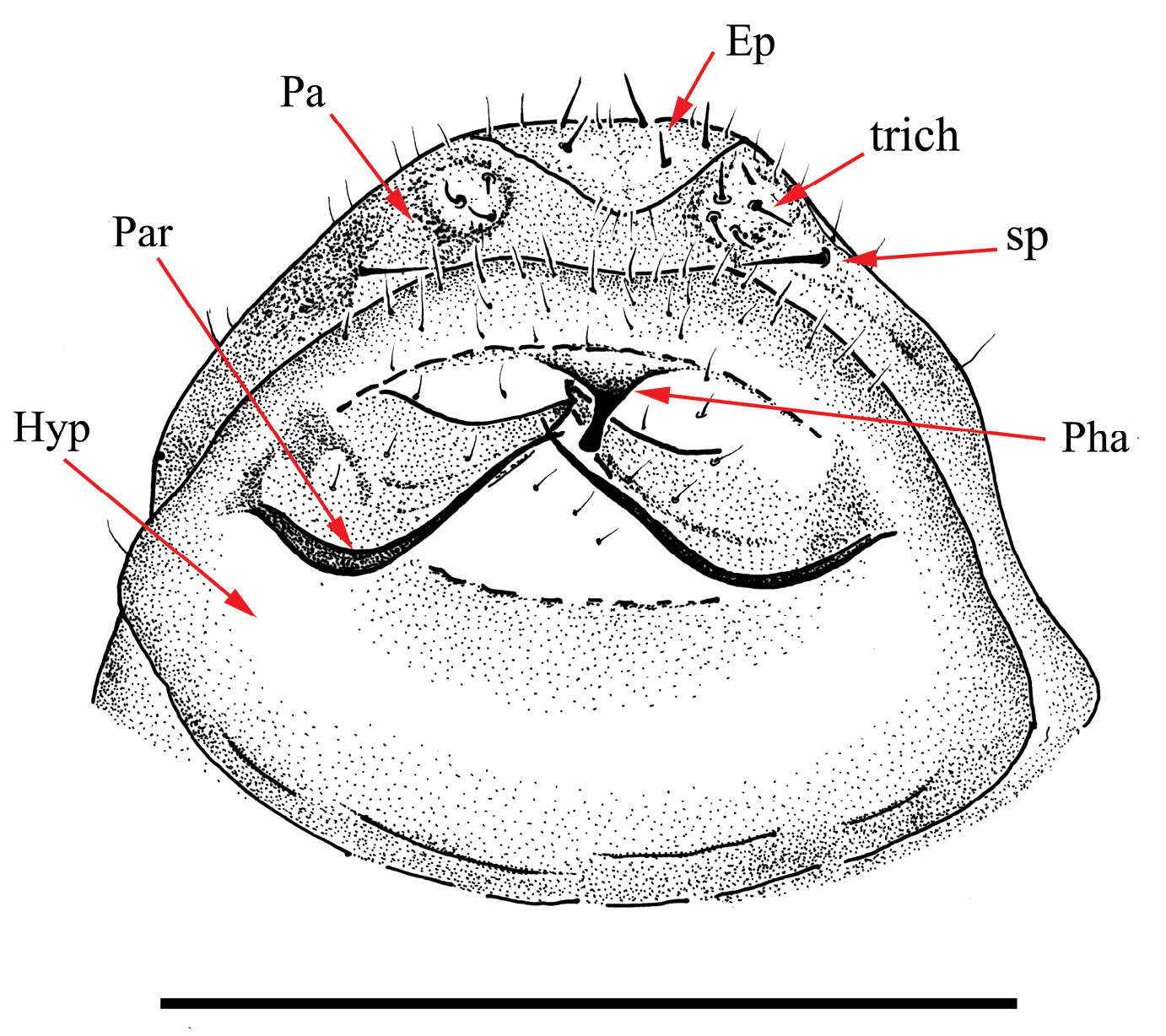
Figure 9.Drawing of aedeagus of Libanopsyllipsocus alexanderasnitsyni gen. et sp. n., holotype, male; Ep = epiproct, Hyp = hypandrium, Par = paraproct, par = paramers, Pha = phallosome, sp = anal spine, trich = trichobothria; scale bar = 0.3 mm.