-
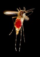
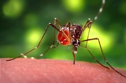
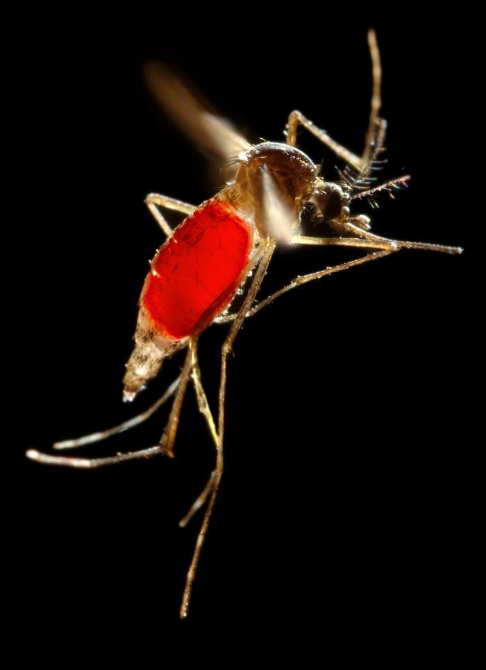
With a newly-obtained fiery red blood meal visible through her now transparent abdomen, the now heavy female Aedes aegypti mosquito takes flight as she leaves her hosts skin surface. In this case, what would normally be an unsuspecting host was actually the CDCs biomedical photographers own hand, which hed offered to the hungry mosquito so that shed alight, and be photographed while feeding. After having filled with blood, the abdomen became distended, stretching the exterior exoskeletal surface, causing it to become transparent, and allowed the collecting blood to become visible as an enlarging intra-abdominal red mass. The wings seem to be working overtime in order to keep her aloft.Created: 2006
-
With a newly-obtained fiery red blood meal visible through her transparent abdomen, the now heavy female Aedes aegypti mosquito took flight as she left her hosts skin surface. In this case, what would normally be an unsuspecting host was actually the CDCs biomedical photographers own hand, which hed offered to the hungry mosquito so that shed alight, and be photographed while feeding. As it filled with blood, the abdomen became distended, stretching the exterior exoskeletal surface, thereby, causing it to become transparent, allowing the collecting blood to become visible as an enlarging intra-abdominal red mass. The wings seem to be working overtime in order to keep her aloft.Created: 2006
-
This 2006 image depicted a female Aedes aegypti mosquito as she was completing the activity of obtaining a blood-meal from a human host through the fascicle of her feeding organ known as the proboscis. Note the droplet of host blood remaining on the tip of her proboscis after shed extracted the feeding organ from the skin surface. In this case, what would normally be an unsuspecting host was actually the CDCs biomedical photographers own hand, which hed offered to the hungry mosquito so that shed alight, and be photographed while feeding. As it filled with blood, the abdomen became distended, stretching the exterior exoskeletal surface, thereby, causing it to become transparent, allowing the collecting blood to become visible as an enlarging intra-abdominal red mass.Created: 2006
-
This 2006 image depicted a female Aedes aegypti mosquito as she was obtaining a blood-meal from a human host through her fascicle, which after penetrating the host's skin, had reddened in color, reflecting the bloods coloration through this tubular structure. In this case, what would normally be an unsuspecting host was actually the CDCs biomedical photographers own hand, which hed offered to the hungry mosquito so that shed alight, and be photographed while feeding. As it filled with blood, the abdomen became distended, stretching the exterior exoskeletal surface, thereby, causing it to become transparent, allowing the collecting blood to become visible as an enlarging intra-abdominal red mass, as is the case in PHIL# 9175, and 9176.Created: 2006
-
This 2006 image depicted a female Aedes aegypti mosquito as she was obtaining a blood-meal from a human host through her fascicle, which after penetrating the host's skin, has reddened in color, reflecting the bloods coloration through this tubular structure. In this case, what would normally be an unsuspecting host was actually the CDCs biomedical photographers own hand, which hed offered to the hungry mosquito so that shed alight, and be photographed while feeding. As it filled with blood, the abdomen became distended, stretching the exterior exoskeletal surface, thereby, causing it to become transparent, allowing the collecting blood to become visible as an enlarging intra-abdominal red mass.Created: 2006
-
This 2006 image depicted a female Aedes aegypti mosquito as she was obtaining a blood-meal from a human host through her fascicle, which had penetrated the host skin, was reddening in color, reflecting the bloods coloration through this tubular structure. In this case, what would normally be an unsuspecting host was actually the CDCs biomedical photographers own hand, which hed offered to the hungry mosquito so that shed alight, and be photographed while feeding. As it filled with blood, the abdomen became distended, stretching the exterior exoskeletal surface, thereby, causing it to become transparent, allowing the collecting blood to become visible as an enlarging intra-abdominal red mass.Created: 2006
-
This 2006 image depicted a female Aedes aegypti mosquito as she was obtaining a blood-meal from a human host through her fascicle, which being transparent, reflected the bloods red color. In this case, what would normally be an unsuspecting host was actually the CDCs biomedical photographers own hand, which hed offered to the hungry mosquito so that shed alight, and be photographed while feeding. As it filled with blood, the abdomen became distended, thereby, stretching the exterior exoskeletal surface, causing it to become transparent, and allowing the collecting blood to become visible as an enlarging intra-abdominal red mass, as is the case in PHIL# 9175, and 9176.Created: 2006
-
This 2006 image depicted a female Aedes aegypti mosquito as she was obtaining a blood-meal from a human host through her fascicle, which being transparent, reflected the bloods red color. In this case, what would normally be an unsuspecting host was actually the CDCs biomedical photographers own hand, which hed offered to the hungry mosquito so that shed alight, and be photographed while feeding. As it filled with blood, the abdomen became distended, stretching the exterior exoskeletal surface, thereby, causing it to become transparent, allowing the collecting blood to become visible as an enlarging intra-abdominal red mass.Created: 2006
-
This 2006 image depicted a female Aedes aegypti mosquito as she was completing the activity of obtaining a blood-meal from a human host through her fascicle, here red in color due to the blood contained therein, which shed begun to resheath in her labium. Both structures are part of her feeding organ known as the proboscis. In this case, what would normally be an unsuspecting host was actually the CDCs biomedical photographers own hand, which hed offered to the hungry mosquito so that shed alight, and be photographed while feeding. As it filled with blood, the abdomen became distended, stretching the exterior exoskeletal surface, thereby, causing it to become transparent, allowing the collecting blood to become visible as an enlarging intra-abdominal red mass, as is the case in PHIL# 9175, and 9176.Created: 2006
-
This 2006 image depicted a female Aedes aegypti mosquito as she was completing the activity of obtaining a blood-meal from a human host through her fascicle, which shed begun to resheath in her labium. Both structures are part of her feeding organ known as the proboscis. In this case, what would normally be an unsuspecting host was actually the CDCs biomedical photographers own hand, which hed offered to the hungry mosquito so that shed alight, and be photographed while feeding. After it filled with blood, the abdomen became distended, stretching the exterior exoskeletal surface, thereby, causing it to become transparent, allowing the collecting blood to become visible as an enlarging intra-abdominal red mass.Created: 2006
-
This 2006 image depicted a female Aedes aegypti mosquito as she was obtaining a blood-meal from a human host through her fascicle, which had penetrated the host skin, was reddening in color, reflecting the bloods coloration through this tubular structure. In this case, what would normally be an unsuspecting host was actually the CDCs biomedical photographers own hand, which hed offered to the hungry mosquito so that shed alight, and be photographed while feeding. As it filled with blood, the abdomen became distended, stretching the exterior exoskeletal surface, thereby, causing it to become transparent, allowing the collecting blood to become visible as an enlarging intra-abdominal red mass, as is the case in PHIL# 9175, and 9176.Created: 2006
-
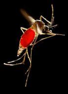
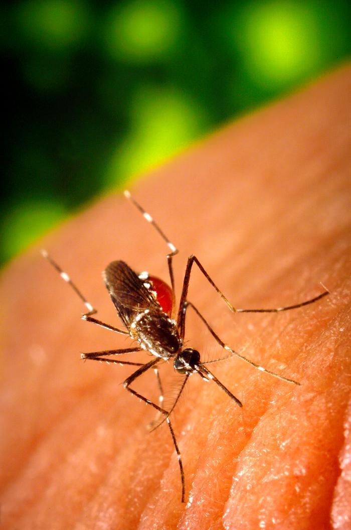
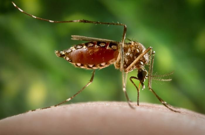
This 2006 image depicted a female Aedes aegypti as she was about to fly off of a hosts skin surface after having obtained her blood-meal. In this case, what would normally be an unsuspecting host was actually the CDCs biomedical photographers own hand, which hed offered to the hungry mosquito so that shed alight, and be photographed while feeding. As it filled with blood, the abdomen became distended, stretching the exterior exoskeletal surface, thereby, causing it to become transparent, allowing the collecting blood to become visible as an enlarging intra-abdominal red mass. The tip of the sharply-pointed fascicle is visible as a yellow tube, which is about to become concealed inside the labium of her proboscis.Created: 2006
-
This 2006 image depicted a female Aedes aegypti mosquito as she was obtaining a blood-meal from a human host through her fascicle, which had penetrated the host's skin, and was reddening in color, reflecting the bloods coloration through this tubular structure. In this case, what would normally be an unsuspecting host was actually the CDCs biomedical photographers own hand, which hed offered to the hungry mosquito so that shed alight, and be photographed while feeding. As it filled with blood, the abdomen became distended, stretching the exterior exoskeletal surface, thereby, causing it to become transparent, allowing the collecting blood to become visible as an enlarging intra-abdominal red mass.Created: 2006
-
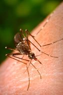
This 2006 photograph depicted a female Aedes aegypti mosquito as she was in the process inserting her fascicle through the skin surface of her host. She then proceeded to obtain a "blood meal", which normally would be from an unsuspecting host, but in this case, the CDC's biomedical photographer, James Gathany, had volunteered his own hand in order to entice the insect to alight, and feed. As it would fill with blood, the abdomen would become distended, thereby, stretching the exterior exoskeletal surface, causing it to become transparent, and allowing the collecting blood to become visible as an enlarging red mass. See PHIL # #8923, for this mosquito's appearance in its abdominally-distended state.Created: 2006
-
This 2006 photograph depicted a female Aedes aegypti mosquito as she was in the process of seeking out a penetrable site on the skin surface of its host. She'd then proceed to obtain a "blood meal", which normally would be from an unsuspecting host, but in this case, the CDC's biomedical photographer, James Gathany, had volunteered his own hand in order to entice the insect to alight, and feed. As it would fill with blood, the abdomen would become distended, thereby, stretching the exterior exoskeletal surface, causing it to become transparent, and allowing the collecting blood to become visible as an enlarging red mass. See PHIL # #8923, for this mosquito's appearance in its abdominally-distended state.Created: 2006
-
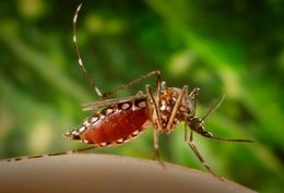
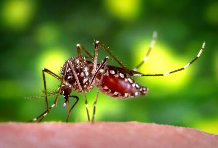
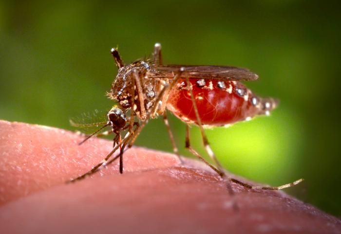
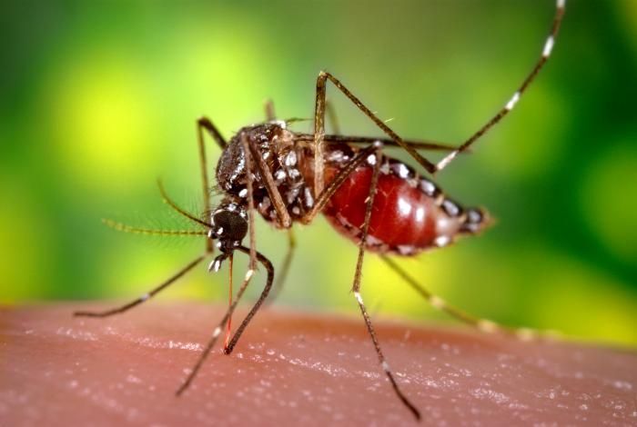
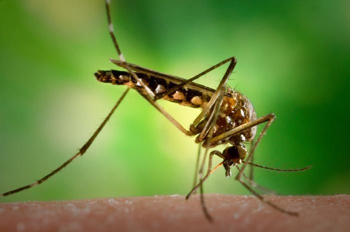
Having finished the ingestion of her blood meal, this 2006 photograph depicted a female Aedes aegypti mosquito as she was in the process of flying from its human host. See PHIL# 8923 - 8933, depicted her in various feeding phases. Normally, this female would be obtaining her blood from an unsuspecting host, but in this case, the CDC's biomedical photographer, James Gathany, had volunteered his own hand in order to entice the insect to alight, and feed. Note that her abdomen had become distended due to the fact that her stomach now filled with her blood meal, and how the proboscis' labial sheath was now pulled up, pointing forward, while no longer was the fascicle inserted in the host's skin.Created: 2006
-
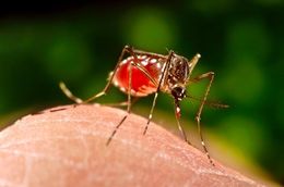
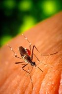
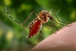
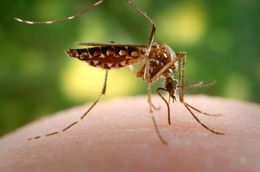
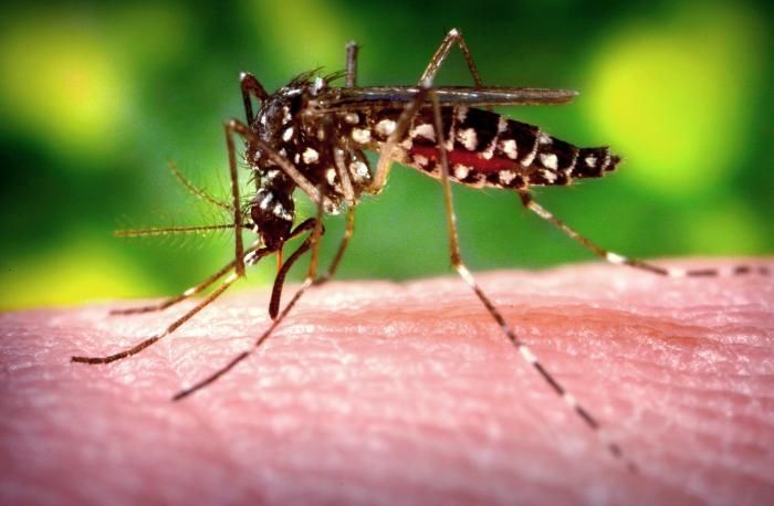
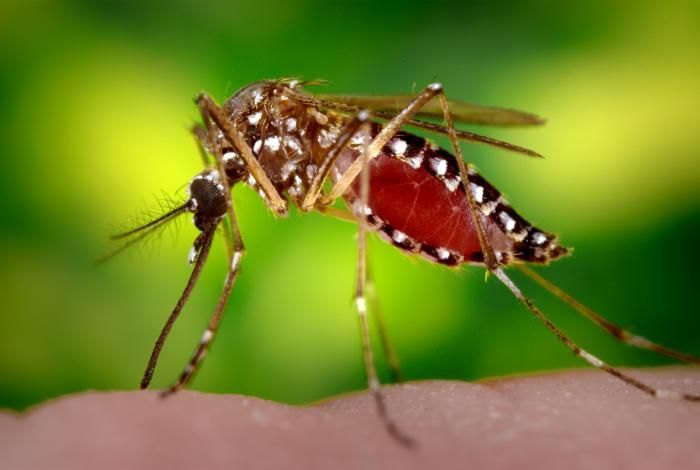
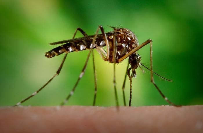
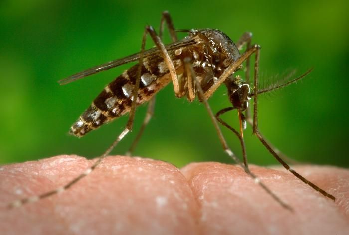
This 2006 photograph depicted a female Aedes aegypti mosquito as she was in the process of acquiring a blood meal from its human host, after having penetrated the skin surface with the sharply-pointed "fascicle". Normally, this female would be obtaining her blood from an unsuspecting host, but in this case, the CDC's biomedical photographer, James Gathany, had volunteered his own hand in order to entice the insect to alight, and feed. Note that her abdomen had become distended as her stomach was filling with her blood meal, and how the proboscis' labial sheath was in its retracted, pulled back configuration, exposing the inserted, sharp fascicle, which had turned red, as the blood was passing up the straw-like apparatus.Created: 2006
-
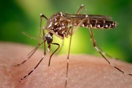
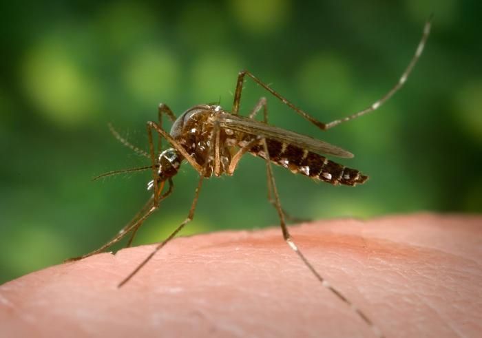
This 2006 photograph depicted a female Aedes aegypti mosquito as she was in the process of acquiring a blood meal from its human host, after having penetrated the skin surface with the sharply-pointed "fascicle". Normally, this female would be obtaining her blood from an unsuspecting host, but in this case, the CDC's biomedical photographer, James Gathany, had volunteered his own hand in order to entice the insect to alight, and feed. Note that her abdomen is beginning to fill with her blood meal, and will continue to fill, becoming distended as it appears in PHIL #8923, and how the proboscis' labial sheath was in its retracted, pulled back configuration, exposing the inserted, sharp fascicle, which had turned red, as the blood was passing up the straw-like needle-sharp apparatus.Created: 2006
-
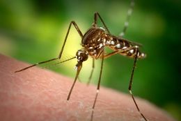
This 2006 photograph depicted a female Aedes aegypti mosquito as she was in the process of beginning the process of acquiring a blood meal from its human host, after having penetrated the skin surface with the sharply-pointed "fascicle". Normally, this female would be obtaining her blood from an unsuspecting host, but in this case, the CDC's biomedical photographer, James Gathany, had volunteered his own hand in order to entice the insect to alight, and feed. As it would fill with blood, the abdomen would become distended, thereby, stretching the exterior exoskeletal surface, causing it to become transparent, and allowing the collecting blood to become visible as an enlarging red mass. See PHIL # #8923, for this mosquito's appearance in its abdominally-distended state.Created: 2006
-
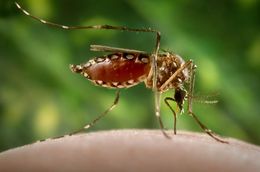
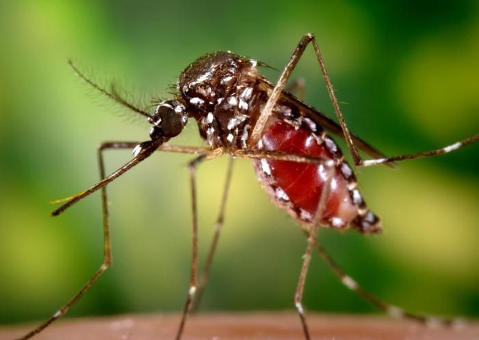
This 2006 photograph depicted a female Aedes aegypti mosquito as she was in the process of acquiring a blood meal from its human host, after having penetrated the skin surface with the sharply-pointed "fascicle". Normally, this female would be obtaining her blood from an unsuspecting host, but in this case, the CDC's biomedical photographer, James Gathany, had volunteered his own hand in order to entice the insect to alight, and feed. As it would fill with blood, the abdomen would become distended, thereby, stretching the exterior exoskeletal surface, thereby, causing it to become transparent, and allowing the collecting blood to become visible as an enlarging red mass. See PHIL # #8923, for this mosquito's appearance in its abdominally-distended state. Note the positioning of the head, and legs, which are closer to the host skin surface, as blood is about to be ingested through the fascicle.Created: 2006
-
This 2006 photograph depicted a female Aedes aegypti mosquito as she was in the process of beginning the process of acquiring a blood meal from its human host, after having penetrated the skin surface with the sharply-pointed "fascicle". Normally, this female would be obtaining her blood from an unsuspecting host, but in this case, the CDC's biomedical photographer, James Gathany, had volunteered his own hand in order to entice the insect to alight, and feed. As it would fill with blood, the abdomen would become distended, thereby, stretching the exterior exoskeletal surface, thereby, causing it to become transparent, and allowing the collecting blood to become visible as an enlarging red mass. See PHIL # #8923, for this mosquito's appearance in its abdominally-distended state.Created: 2006