-
Marko Lukić, Céline Houssin, Louis Deharveng
Zookeys
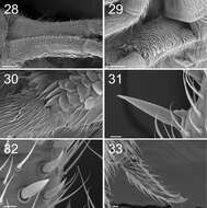
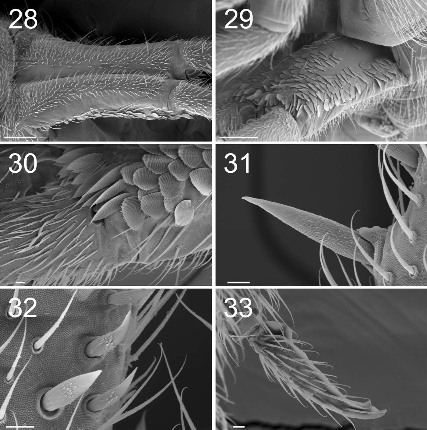
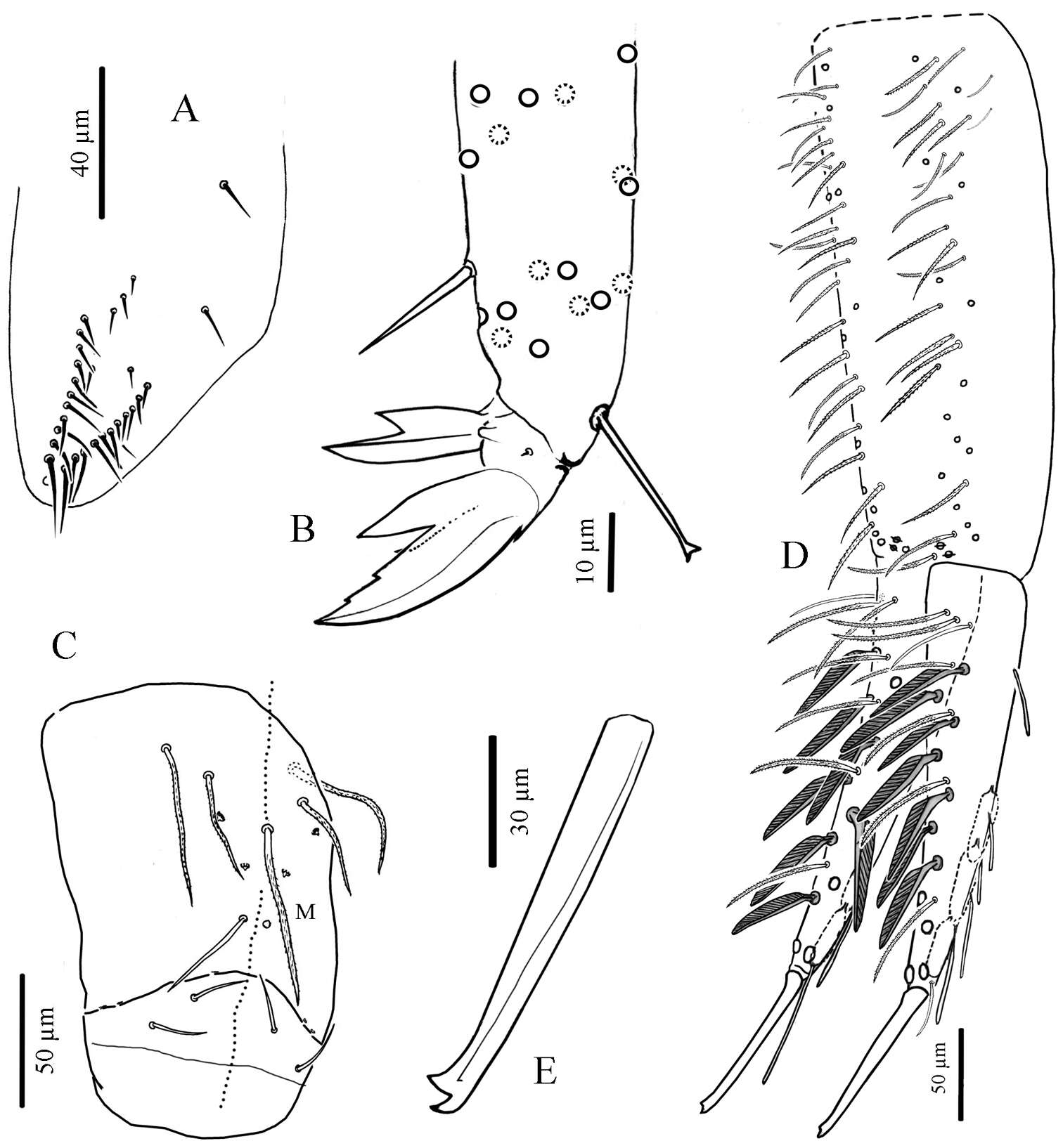
Figures 28–33. Tritomurus veles sp. n. (SEM). 28 Manubrium in dorsal view (scale 100 μm) 29 Manubrium in ventral view (scale 100 μm) 30 Manubrium ventro-distally and dens ventro-basally (scale 10 μm) 31, 32 Dental spines (scale 10 μm) 33 Mucro (scale 10 μm).
-
Asker, Akershus, Norge
-
Asker, Akershus, Norge
-
Asker, Akershus, Norge
-
Xiang-Qun Yuan, Zhi-Xiang Pan
Zookeys
Figure 35.dorsal chaetotaxy of Abd. I–III of Sinella triseta sp. n.
-
Sopark Jantarit, Chutamas Satasook, Louis Deharveng
Zookeys
Figure 8.Cyphoderus khaochakanus sp. n. A trochanteral organ B claw and distal part of tibiotarsus III C posterior face of the ventral tube D furca; feathered chaetae in lateral view, only one of the two vanes attached to the rachis is visible E mucro.
-
Marko Lukić, Céline Houssin, Louis Deharveng
Zookeys
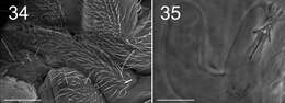
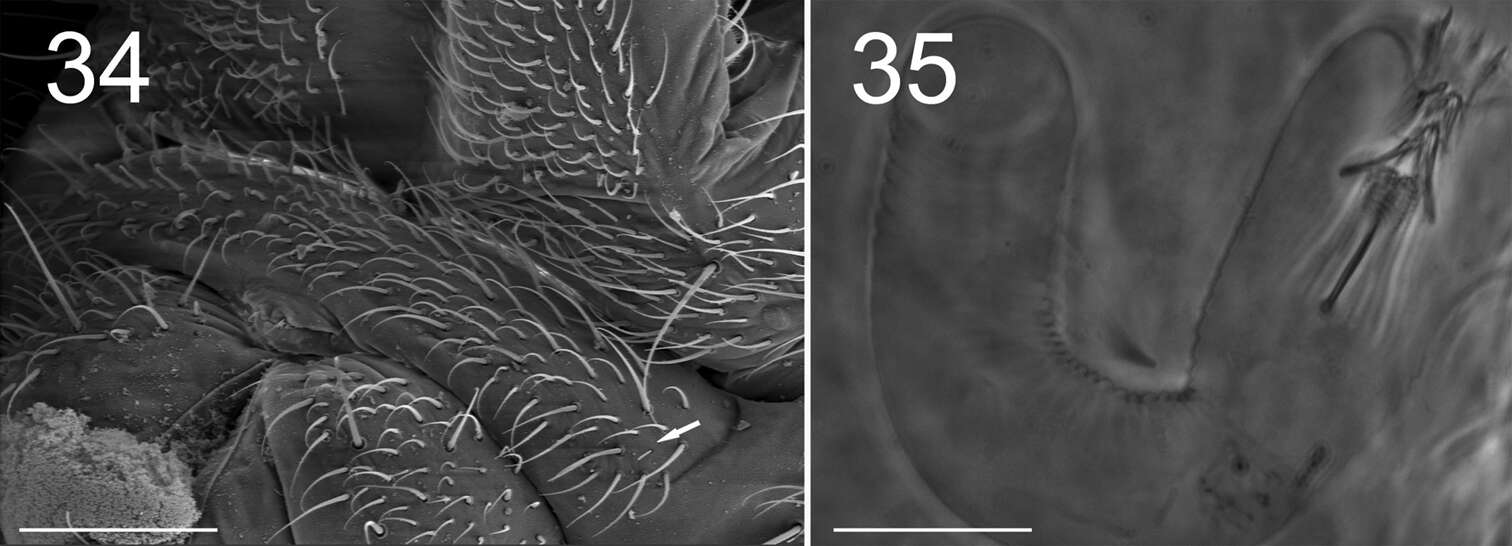
Figures 34–35. Tritomurus veles sp. n. (34, SEM; 35, optical microscope). 34 Sternite of Abd.V with genital plate (scale 100μm); arrow points to minute lateral S-microchaeta 35 Internal parasitic larva (Nematomorpha) (scale 20 µm).
-
Asker, Akershus, Norge
-
Asker, Akershus, Norge
-
Sopark Jantarit, Chutamas Satasook, Louis Deharveng
Zookeys
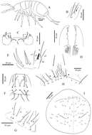
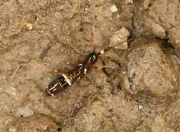
Figure 2.Cyphoderus songkhlaensis sp. n. A habitus B outer maxillary lobe C maxilla head and ventral complex of the labrum D mandible E labial palp: proximal chaetae and external papilla E F labrum, dorsal view G chaetotaxy of labial basis; frontal chaetae H frontal chaetae and pseudopores of head I dorsal chaetotaxy of head.
-
Marko Lukić, Céline Houssin, Louis Deharveng
Zookeys
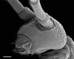
Figure 4. Tritomurus veles sp. n., head in lateral view (SEM, scale 100 μm).
-
Asker, Akershus, Norge
-
Asker, Akershus, Norge
-
Marko Lukić, Céline Houssin, Louis Deharveng
Zookeys
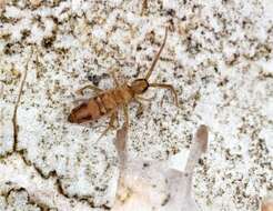
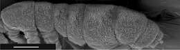
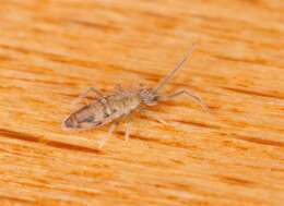
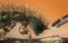
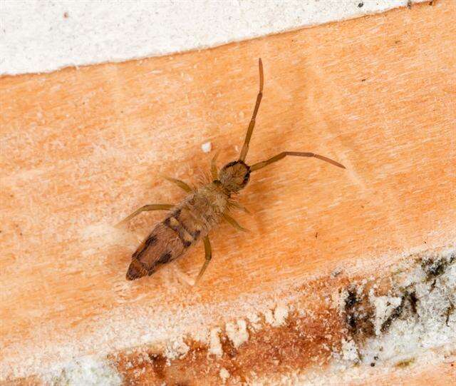
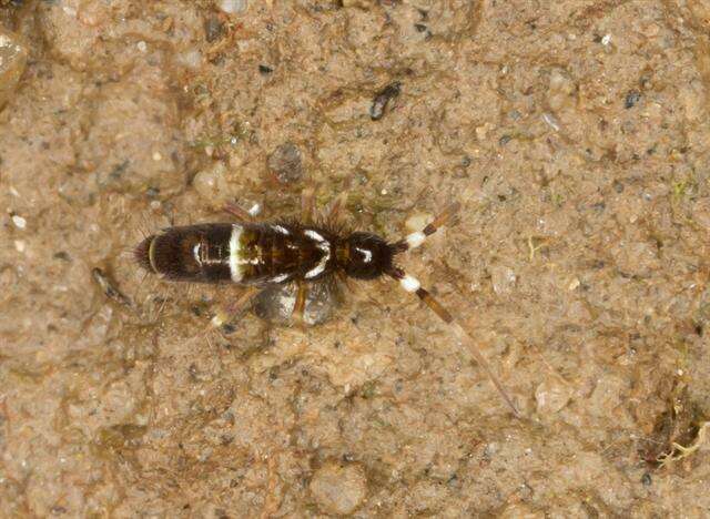
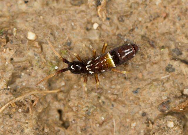
Figure 5. Tritomurus veles sp. n., body (SEM, scale 400 μm).
-
Asker, Akershus, Norge
-
Asker, Akershus, Norge
-
Marko Lukić, Céline Houssin, Louis Deharveng
Zookeys
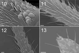
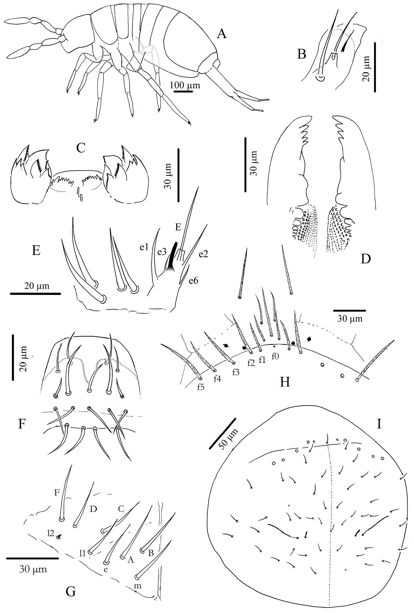
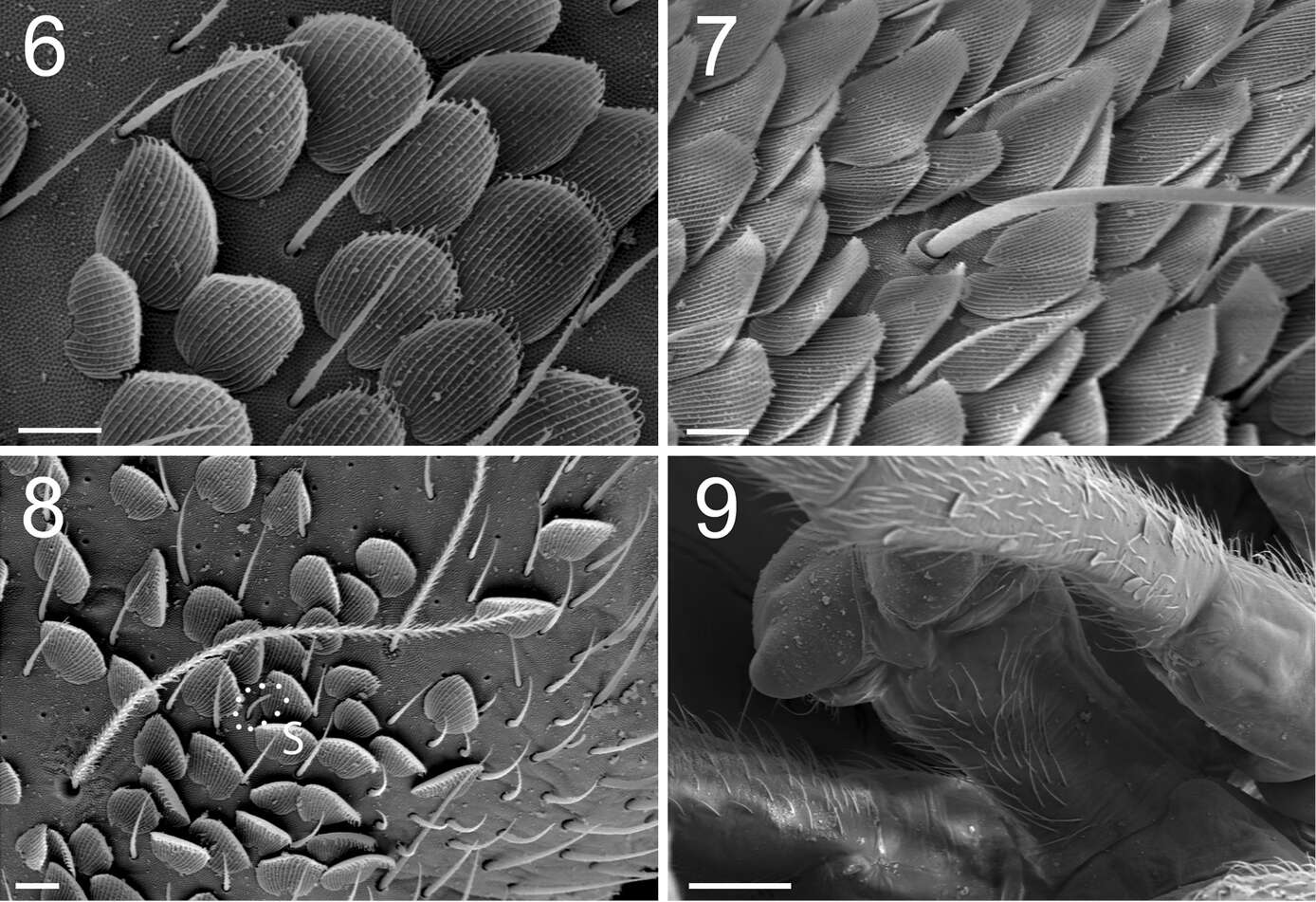
Figures 6–9. Tritomurus veles sp. n. (SEM). 6 Scales and ordinary mesochaetae on Th.II (scale 10 μm) 7 Scales, mesochaetae and macrochaeta on Abd.III (scale 10 µm) 8 Bothriotrichal area of Th.II, illustrating the presence of the four main chaetal types: bothriotricha, mesochaetae, S-chaetae (S) and scales (scale 10 µm) 9 Ventral tube in anterior view (scale 100 µm).
-
Asker, Akershus, Norge
-
Marko Lukić, Céline Houssin, Louis Deharveng
Zookeys
Figures 10–13. Tritomurus veles sp. n. (10–12, SEM; 13, optical microscope) 10 Lateral view of Ant.I, with scales, mesochaetae, and latero-distal short S-microchaetae at left (scale 10 µm) 11 Ant.III proximally (scale 10 μm) (S, S-chaetae) 12 Ant.IV (scale 10 μm) (S, S-chaetae) 13 Apical part of Ant.IV (scale 10 μm).
-
Asker, Akershus, Norge
-
Marko Lukić, Céline Houssin, Louis Deharveng
Zookeys
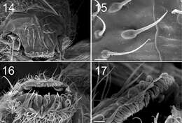
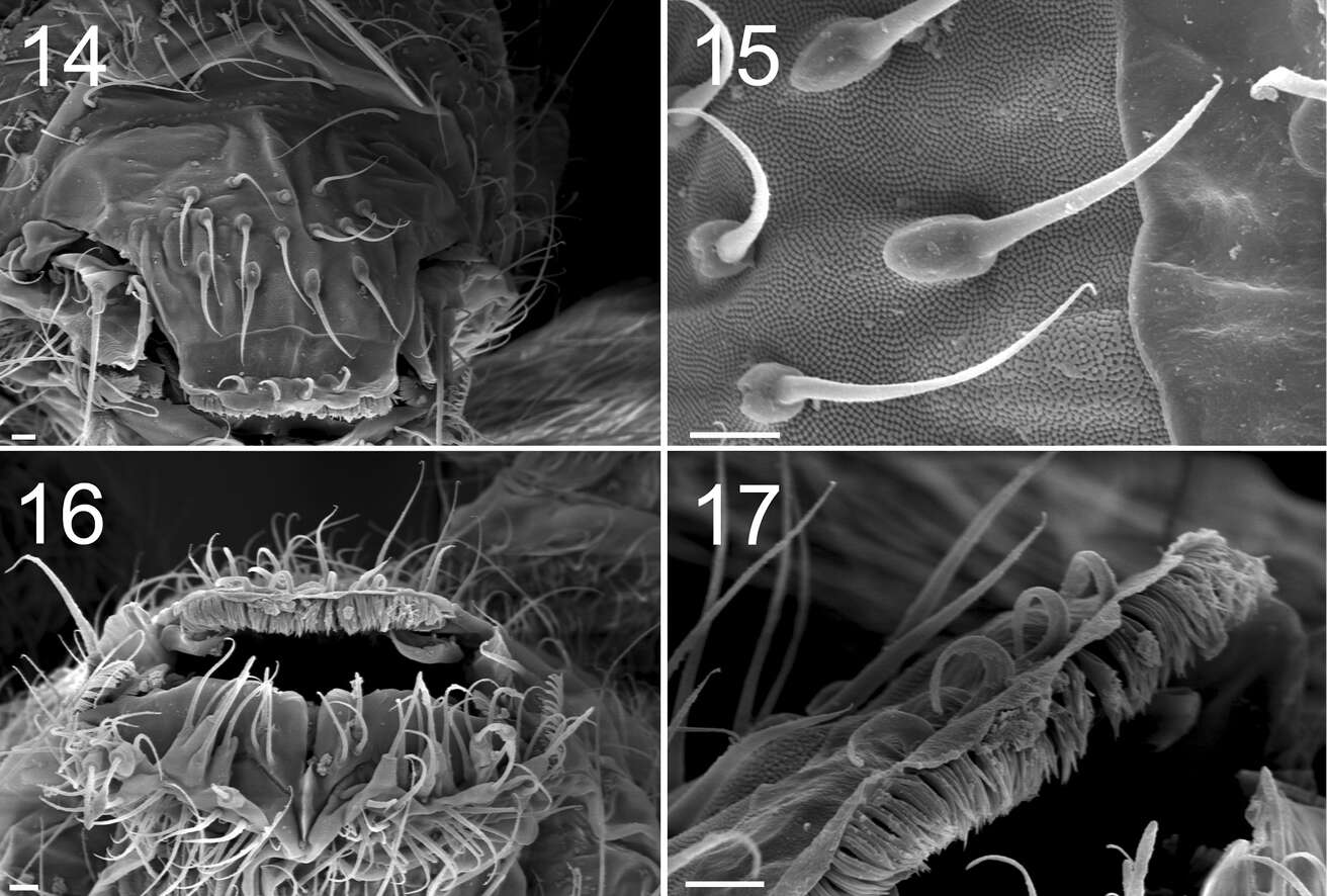
Figures 14–17. Tritomurus veles sp. n. (SEM) 14 Labrum (scale 10 μm) 15 Detail of labral chaetae with swollen socket (scale 10 µm) 16 Mouth (scale 10 μm) 17 ventro-distal brush of labrum (scale 10 μm).
-
Marko Lukić, Céline Houssin, Louis Deharveng
Zookeys
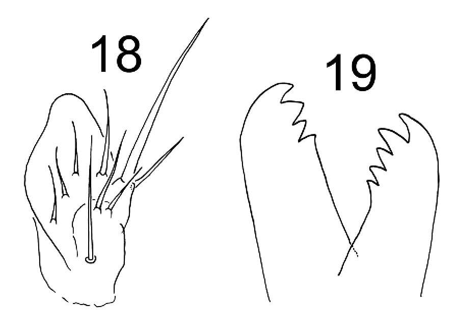
Figures 18–19. Tritomurus veles sp. n. 18 Outer maxillary lobe 19 Left and right mandibles.
-
Marko Lukić, Céline Houssin, Louis Deharveng
Zookeys
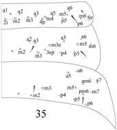
Figure 20. Tritomurus veles sp. n., macrochaetotaxic and bothriotrichal pattern; plain line circles, macrochaetae; dotted line circles, small macrochaetae or large mesochaetae; lat, lateral group of Abd.IV; X, bothriotricha) (scale 400 μm).
-
Marko Lukić, Céline Houssin, Louis Deharveng
Zookeys
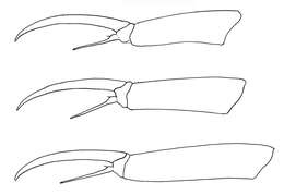
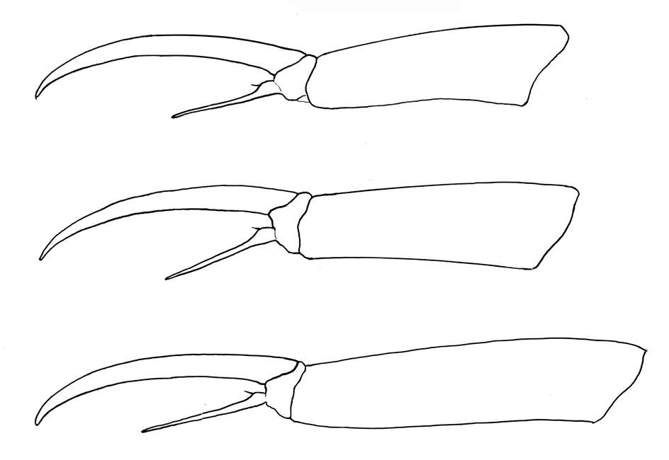
Figure 21. Tritomurus veles sp. n. Tibiotarsus and claw of legs I, II, III from upper to lower.