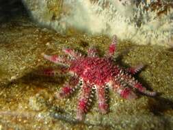
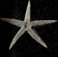
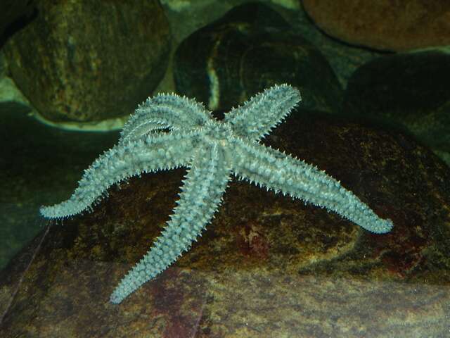
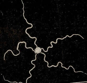
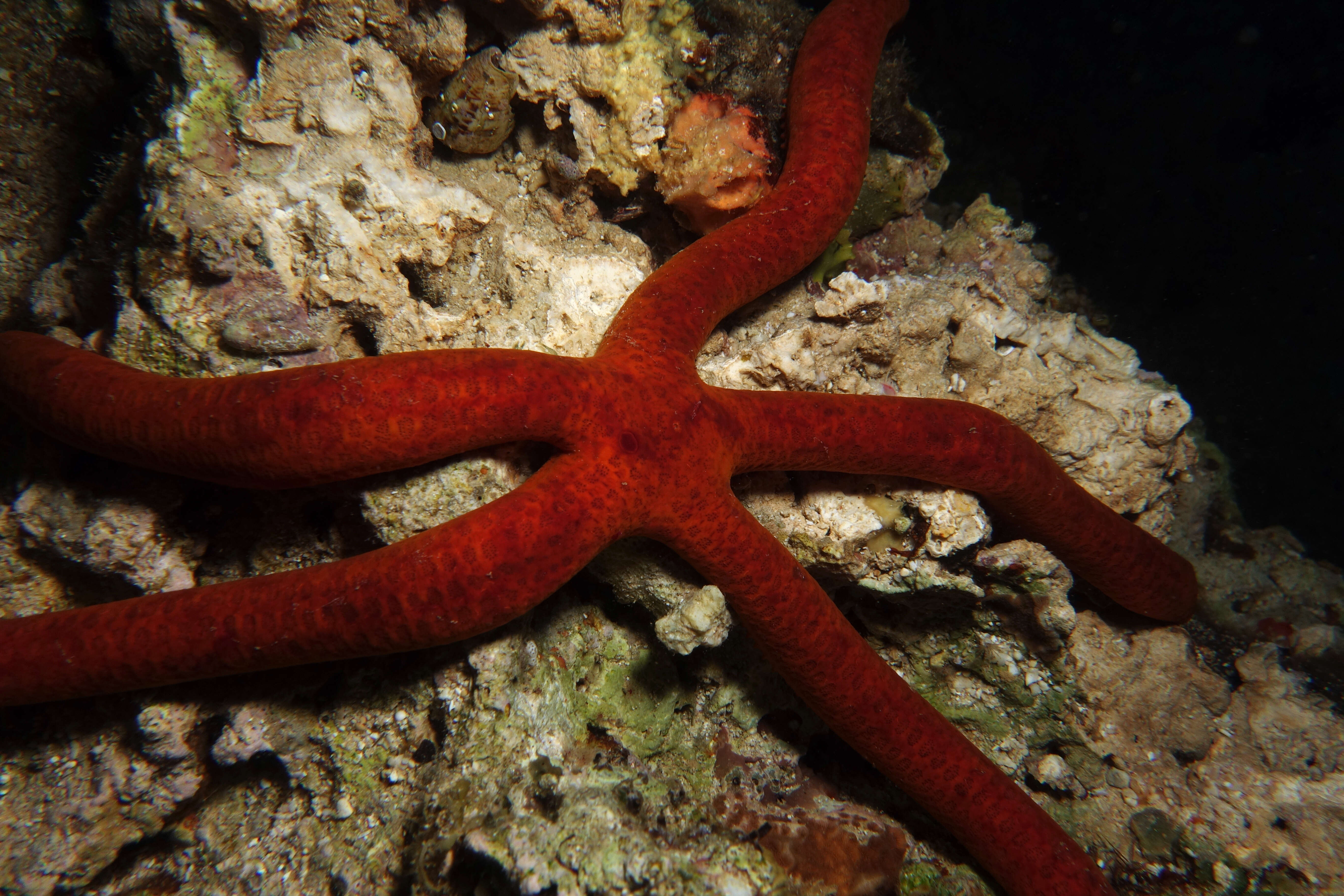
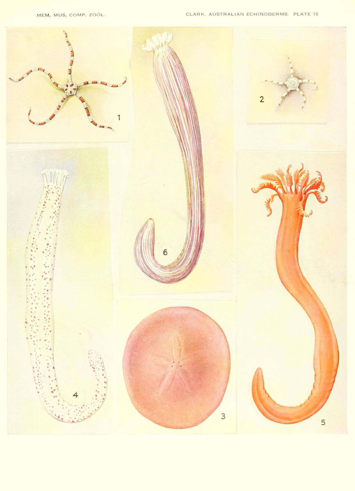
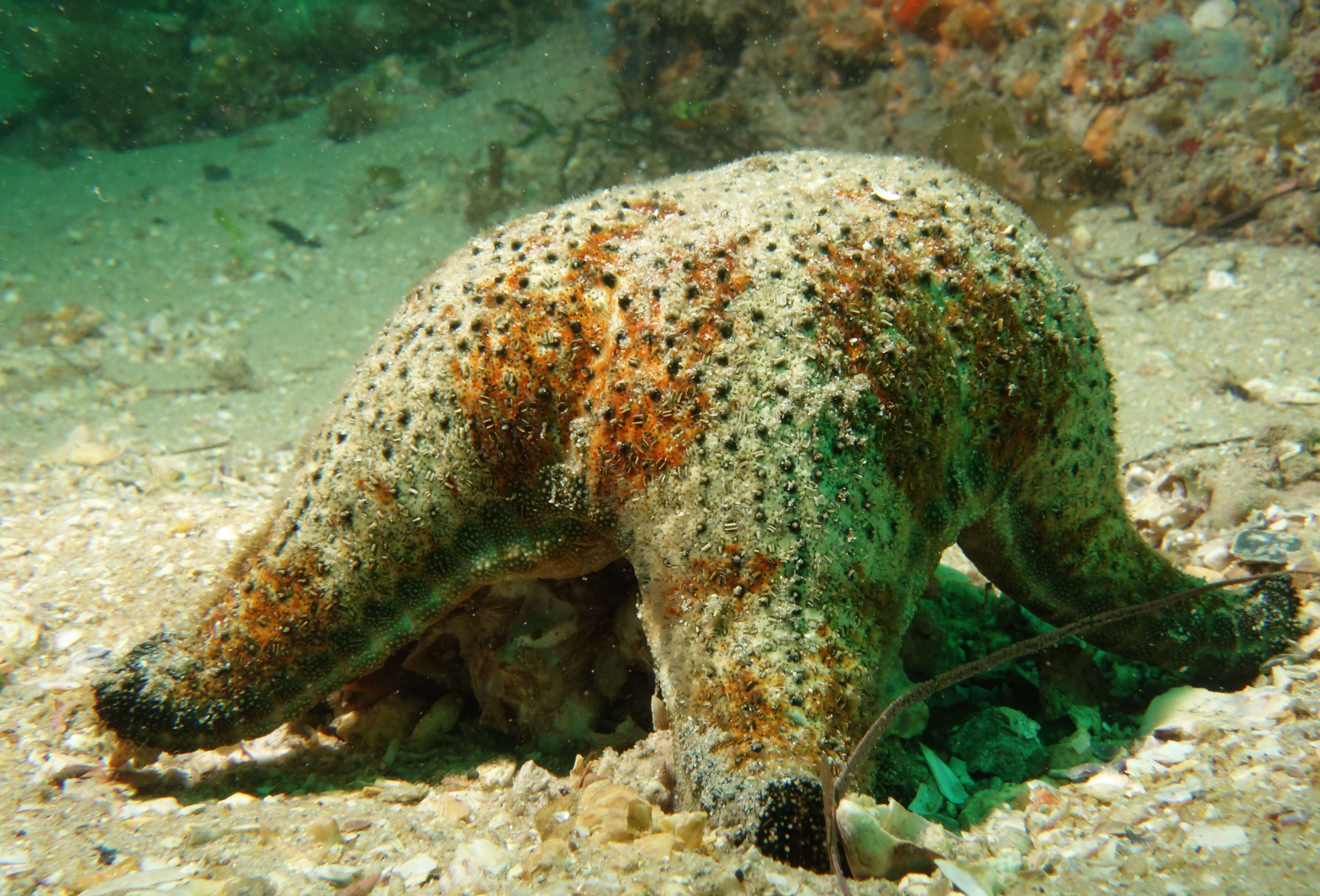
Deep-sea sun star (Rathbunaster californicus) .This photo was taken at a depth of -404.4 meters in Sur Canyon as a part of a deep-sea coral expedition conducted by NMFS aboard the R/V Shimida in December, 2010. Photo taken Dec. 21, 2010, Location: Point Sur, Sur Canyon.Kevin L. Stierhoff / NOAA SWFSC From: SIMoN
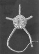
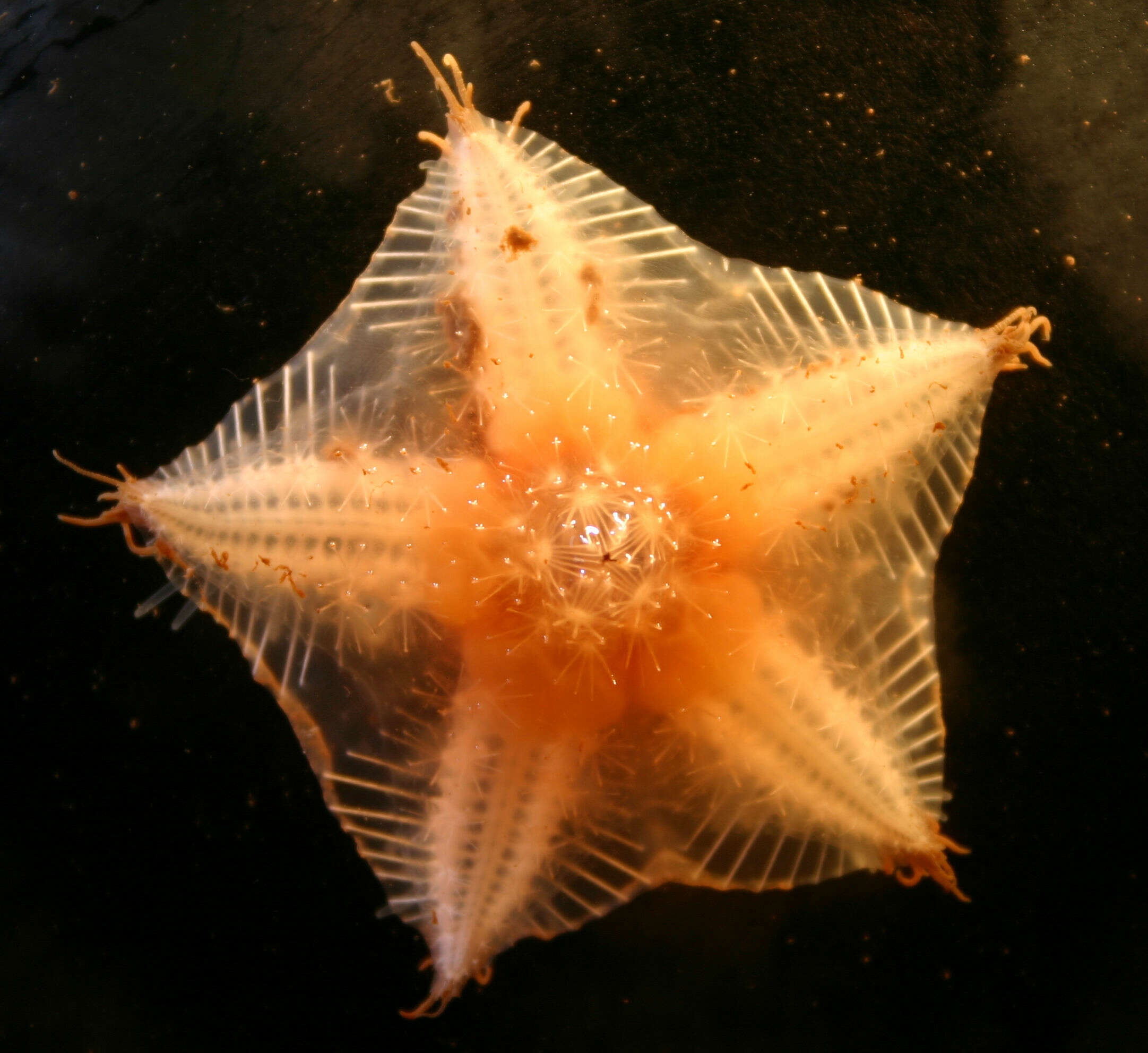
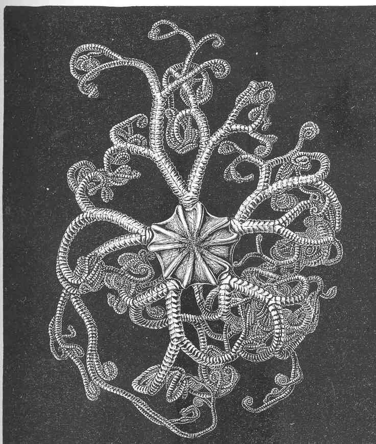
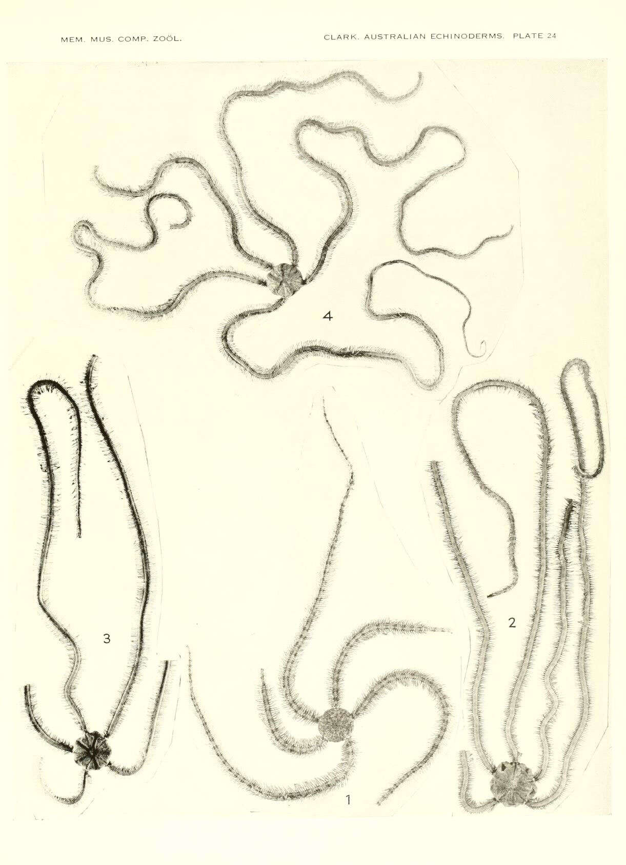
Indian Deep-Sea Starfishes. Pontaster hispidus, from the Laccadive Sea, 1000 fathoms. Back view, showing the plots of papules at the bases of the rays. Only one ray is represented at full length
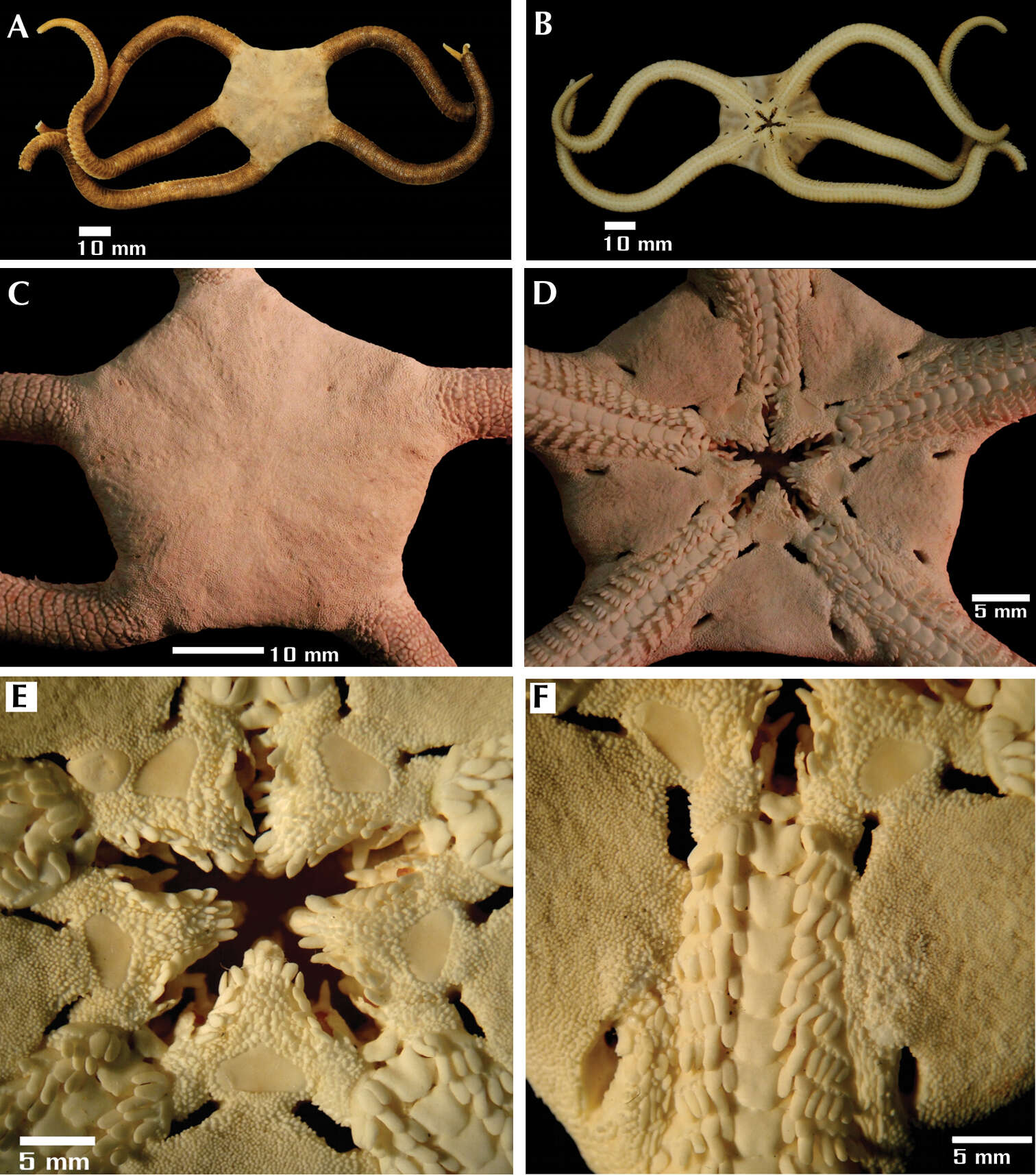
Masanori Okanishi, Timothy D. O’Hara, Toshihiko Fujita
Zookeys
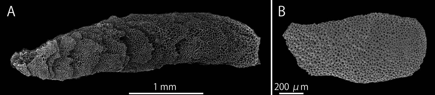
Figure 1.SEM photographs of radial shields of Asteroschema tubiferum (NSMT E-2110) (A) and Squamophis albozosteres sp. n., paratype(MV F-162658) (B). A multi-layered radial shield B single-layered radial shields. The left side of the images are towards the center of the disc and the right side towards the disc margin.
Anne I. Gondim, Carmen Alonso, Thelma L. P. Dias, Cynthia L. C. Manso, Martin L. Christoffersen
Zookeys
Figure 2.Species of the families Ophiomyxidae (A–E) and Ophiotrichidae (F–J). Ophiomyxa flaccida. A dorsal view, in detail the marginal interradius with a row of large scales B ventral view C jaw D dorsal view of the arm E ventral view of the arm. Ophiothrix (Ophiothrix) angulata F dorsal view, in detail the radial shields G ventral view H jaw I dorsal view of the arms J ventral view of the arms. Scale bar = 1 mm.
Tania Pineda-Enríquez, Francisco A. Solís-Marín, Yuri Hooker, Alfredo Laguarda-Figueras
Zookeys
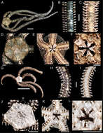
Figure 2.Ophioderma peruana sp. n., holotype (CZA-363). A aboral view B oral view C aboral disc and basal portion of the arms D oral disc and basal portion of the arms E jaws F oral portion of the disc and pair of genital slits.
Rebeca Granja–Fernández, María D. Herrero-Pérezrul, Ramón A. López-Pérez, Luis Hernández, Fabián A. Rodríguez-Zaragoza, Robert Wallace Jones, Rubén Pineda-López
Zookeys
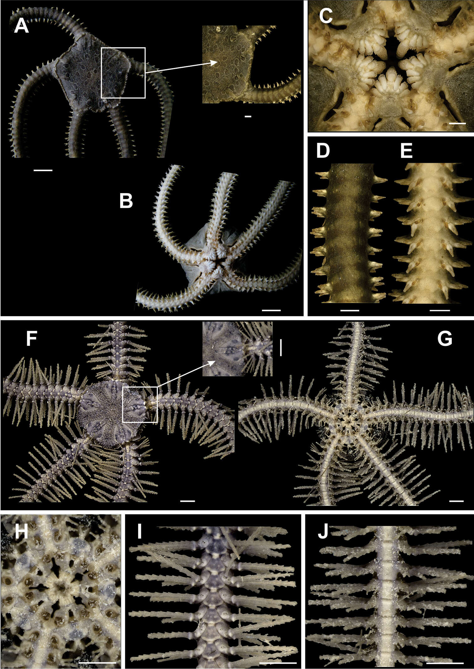
Figure 1.Ophiocnida hispida. A dorsal view. Scale bar = 5 mm B dorsal view of the arm C ventral view of the arm D dorsal view of the disk E ventral view of the disk F jaw. Scale bar = 1 mm. Ophiophragmus papillatus G dorsal view. Scale bar = 5 mm H dorsal view of the arm I ventral view of the arm J dorsal view of the disk (p = papillae around the margin of the disk) K ventral view of the disk L jaw. Scale bar = 1 mm.