-
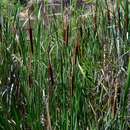
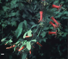
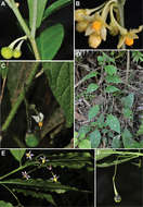
Figure 45.Leaves, flower and fruit of Passiflora mcvaughiana (Porter-Utley & Mondragón 345) Scale bar = 10.0 mm.
-
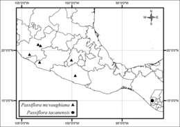
Figure 46.Distribution of Passiflora mcvaughiana and Passiflora tacanensis.
-
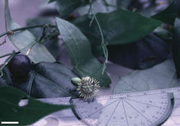
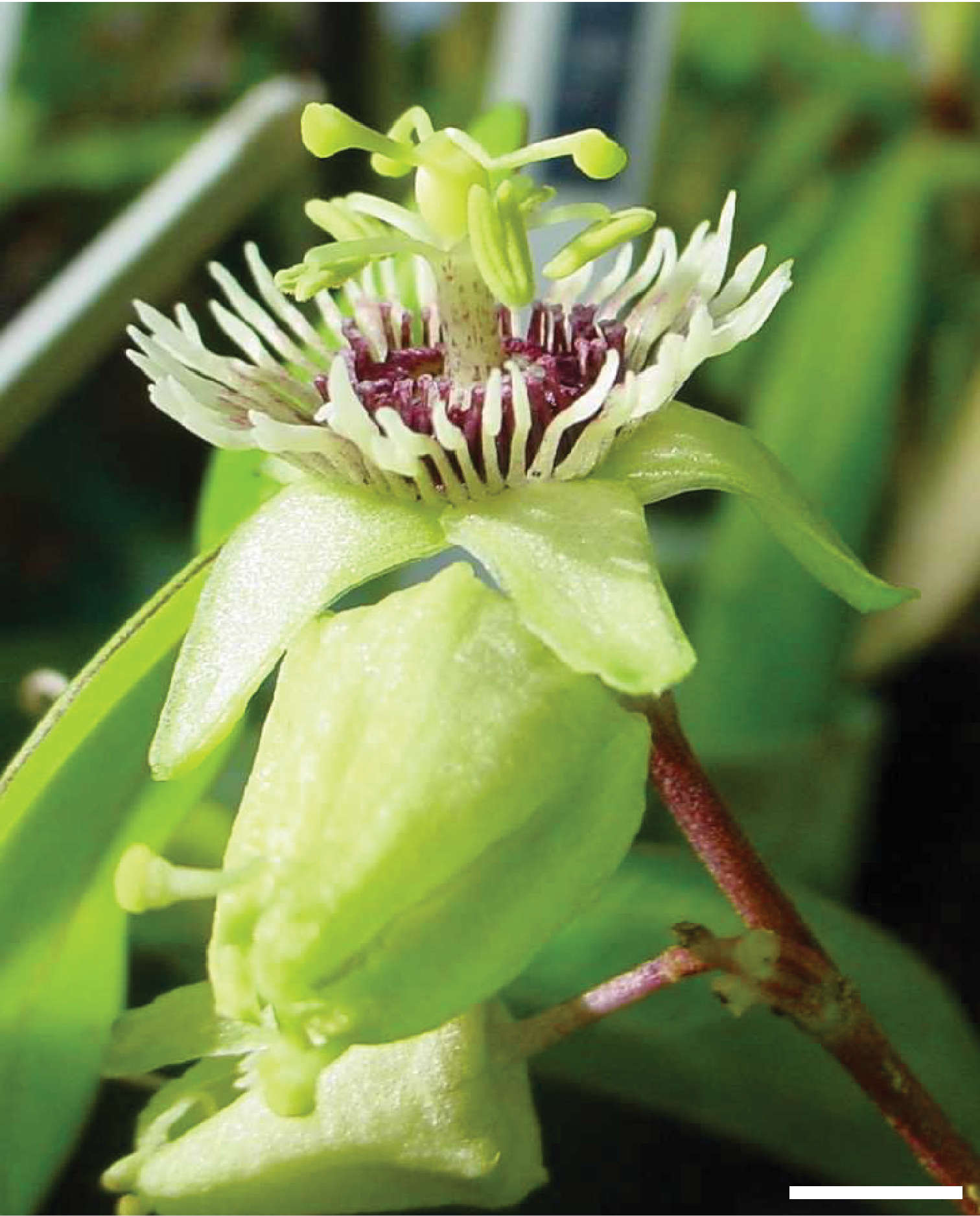
Figure 48.Flower of Passiflora coriacea from Colombia. Scale bar = 5.0 mm. Photo by C. Feuillet.
-
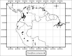
Figure 49.Distribution of Passiflora coriacea.
-
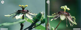
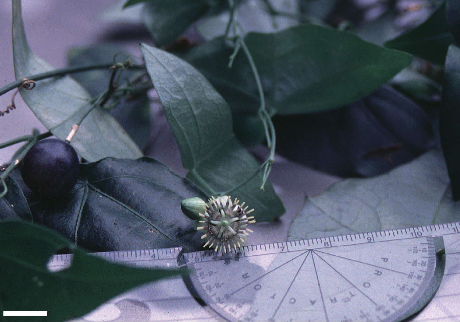
Figure 52.Leaf, flower, inflorescence, and fruit of Passiflora sexocellata from plant growing in greenhouse at Butterfly World, Coconut Creek, Florida. Scale bar = 10 mm.
-
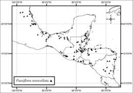
Figure 53.Distribution of Passiflora sexocellata.
-
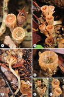
Figure 54.a Flower of Passiflora itzensis (MacDougal 4633) Scale bar = 5.0 mm. Photo by J. M. MacDougal b Flower of Passiflora xiikzodz (MacDougal 4677) Scale bar = 5.0 mm. Photo by J. M. MacDougal.
-
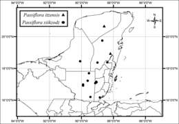
Figure 55.Distribution of Passiflora itzensis and Passiflora xiikzodz.
-
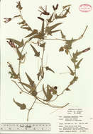
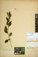
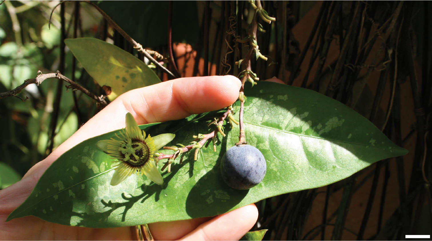
Figure 31.Herbarium specimen of Passiflora lancifolia (G. Proctor 23725).
-
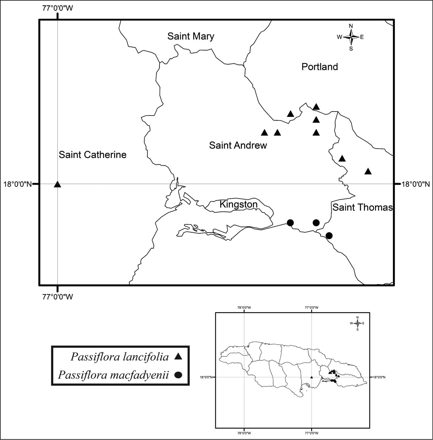
Figure 32.Distribution of Passiflora lancifolia and Passiflora macfadyenii.
-
Figure 32.Distribution of Passiflora lancifolia and Passiflora macfadyenii.
-
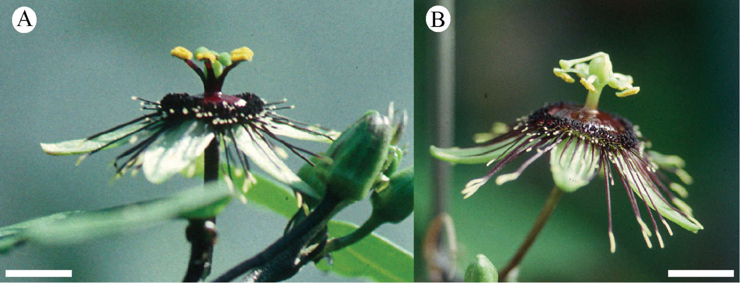
Figure 33.Leaves and flowers of Passiflora macfadyenii (MacDougal 452) Scale bar = 6.0 mm. Photo by J. M. MacDougal.
-
Tiina Särkinen, Paúl Gonzáles, Sandra Knapp
Phytokeys
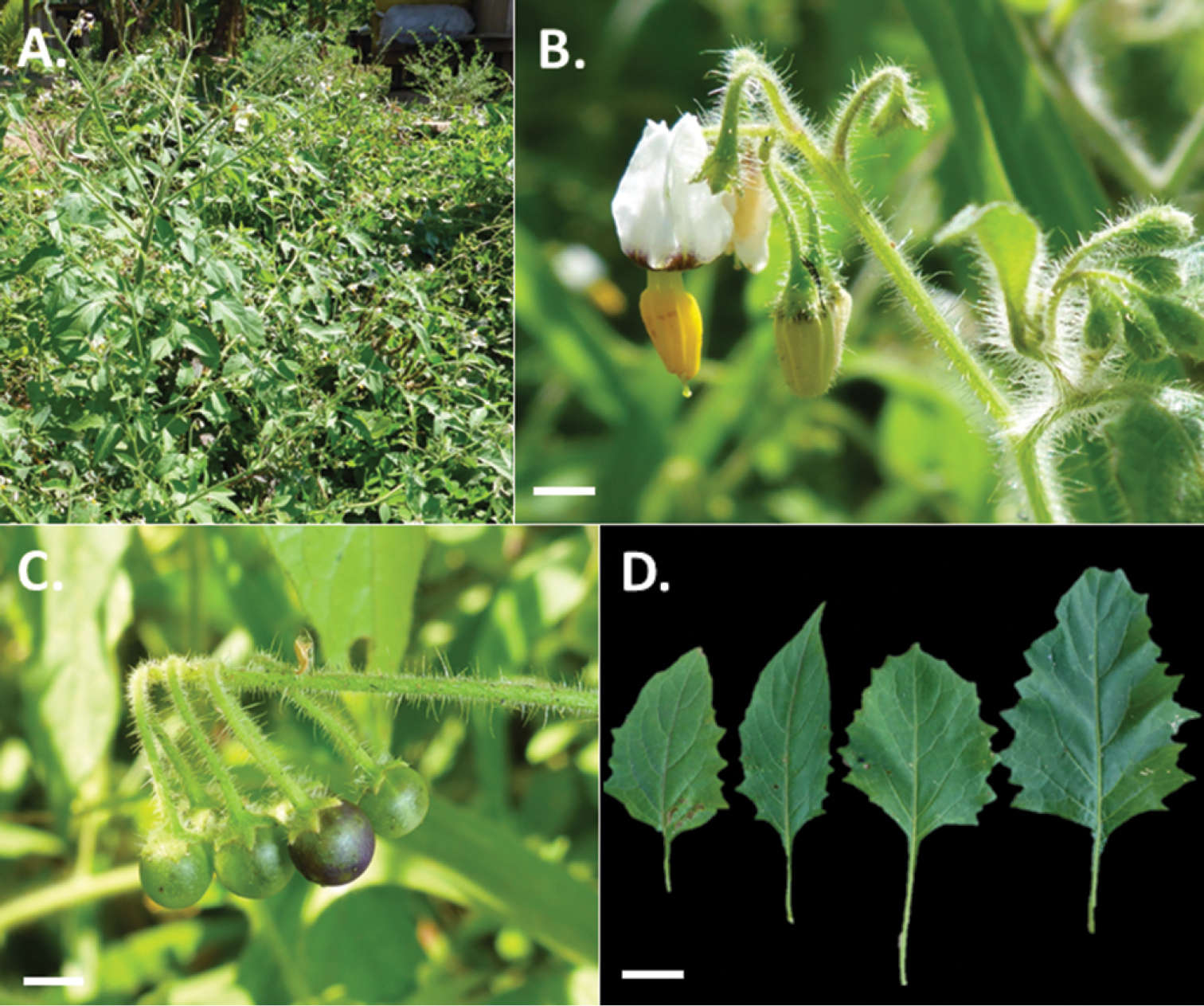
Figure 5.Photos of Solanum arenicola. A Habit B Buds and flowers, showing the dense indumentum of glandular-tipped, multi-cellular hairs throughout C Maturing fruits, showing reflexed pedicels in infrutescence D Leaf size and shape variation present within individuals as observed in the field (A–D Särkinen & Balarezo 4866). Scale bars = 1 mm. All photos by T. Särkinen.
-
Tiina Särkinen, Paúl Gonzáles, Sandra Knapp
Phytokeys
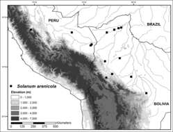
Figure 6.Distribution map of Solanum arenicola in lowlands of central and southern Peru, and northern Bolivia.
-
Shek Shing Mar, Richard M.K. Saunders
Phytokeys
Figure 1.Flower development in Thismia hongkongensis sp. nov. A, B Root system, with young flowering stalk developing (arrowed). C–H Developing flower, photographed over a 17-day period (10th, 14th, 16th, 19th, 23rd and 27th May, respectively) (S.S. Mar 1, HK). I, J Post-fertilization flower, showing abscission of perianth tube. Photos by S.S. Mar.
-
Shek Shing Mar, Richard M.K. Saunders
Phytokeys
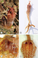
Figure 2.Flower structure in Thismia hongkongensis sp. nov. A Mature flower, showing outer tepals (ot), inner tepals (it) and abscission zone (ab) at the base of the perianth tube. B Entire plant (S.S. Mar 1, HK). C Perianth tube with annulus (a), following removal of the proximal face of the tube, exposing pendent stamens with filament (f), thecae (th), connective (c) and lateral appendage (la) (S.S. Mar 2, HK). D Inner face of perianth tube, showing network patterning and putative nectaries (arrowed) (S.S. Mar 2, HK). Scale bars: A, D = 2 mm; B = 5 mm; C = 1 mm. Photos: A, B S.S. Mar; C, D R.M.K. Saunders.
-
Shek Shing Mar, Richard M.K. Saunders
Phytokeys
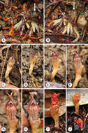
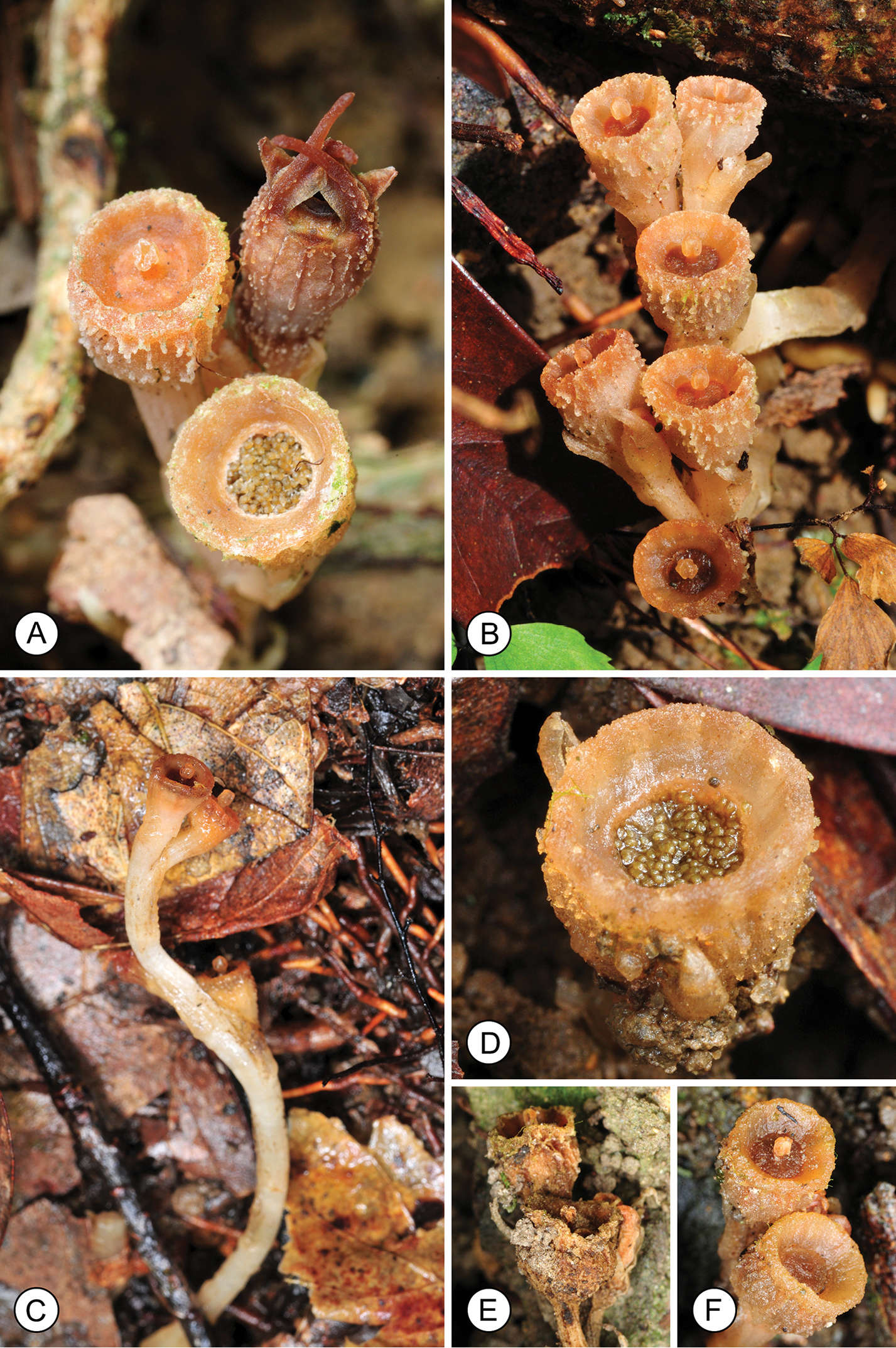
Figure 3.Fruit structure in Thismia hongkongensis sp. nov. A Flower (rear right), immature fruit, shortly after fertilization (left), and mature fruit with exposed seeds (front). B Two fruiting individuals, each with three fruits. C Lateral view of fruiting specimen, illustrating elongated fruit stalk. D Mature fruit with exposed seeds. E Dehydrated fruit. F Rehydrated fruit, after rainfall. Photos by S.S. Mar.
-
Shek Shing Mar, Richard M.K. Saunders
Phytokeys
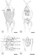
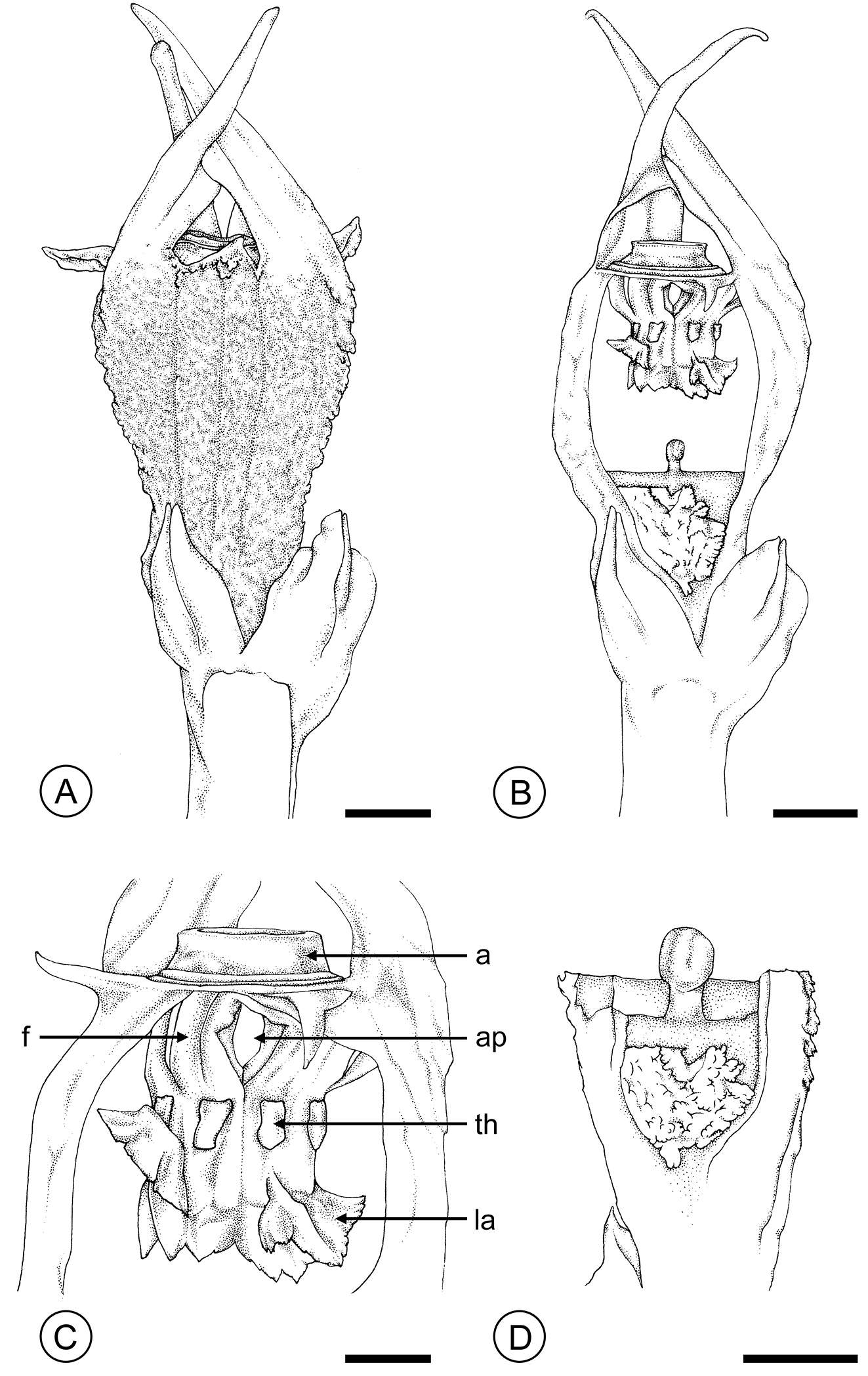
Figure 4.Thismia hongkongensis sp. nov. (S.S. Mar 2, HK). A Entire flower. B Flower with proximal part of perianth tube removed, showing pendent stamens. C Apex of the perianth tube, showing annulus (a) and pendent stamens, with filament (f), thecae (th), lateral appendage (la), and aperture (ap) between filaments. D Longitudinal section through fused carpels. Scale bars: A, B, D = 2 mm; C = 1 mm. Drawings by Caren Pearl Shin.
-
James W. Byng, F. B. Vincent Florens, Cláudia Baider
Phytokeys
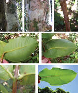
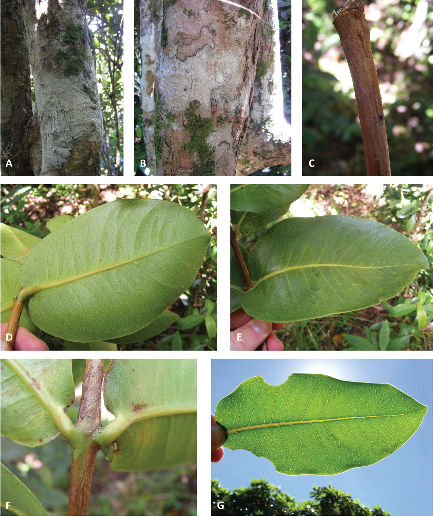
Figure 1.Vegetative characters. A and B bark C close-up of branchlet D lower leaf surface E upper leaf surface F petioles G leaf venation. (A and G Byng 83; B–F Byng 84).
-
James W. Byng, F. B. Vincent Florens, Cláudia Baider
Phytokeys
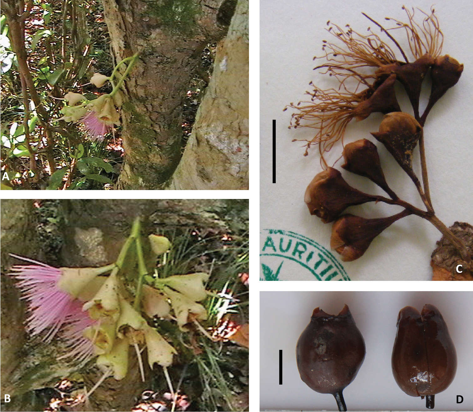
Figure 2.Floral and fruit characters. A and B Sole recorded images of flowering event C Close-up of dried inflorescence D Close-up of two fruits. (A–C D’Argent & Pynee MAU 25014; D D’Argent & K. Pynee MAU 26448; A and B courtesy of Kersley Pynee). Scale bar = 1 cm.
-
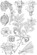
Figure 1.Heliotropium perlmanii Lorence & WL Wagner A Habit B Upper leaf surface, C Inflorecence D Flower, lateral view E Corolla, face view F Flower, longitudinal section showing stamens and gynoecium G Corolla, sectioned to show stamens and indument, H Fruit and calyx, lateral view I Fruit showing 4 carpels. All figures drawn from Perlman & Florence 10052 (US) and photos from Falaise Est Eiao, 11 March 2007 courtesy of J-F Butaud.
-
Figure 2.Heliotropium marchionicum Decne. A Habit B Upper Leaf surface C Inflorescence D Flower, lateral view E Corolla, face view F Flower, longitudinal section showing stamens and gynoecium G Corolla, sectioned to show stamens and indument H Fruit and calyx, lateral view I Fruit showing 4 carpels. Drawn from Perlman 10005 (US) and photos from Nuku Hiva, 24 February 2007 [A], Mercier 1847 (US) and photos from Nuku Hiva, 24 February 2007 courtesy of J-F Butaud [B–I].
-
Sandra Knapp, João Renato Stehmann, Leandro L. Giacomin
Phytokeys
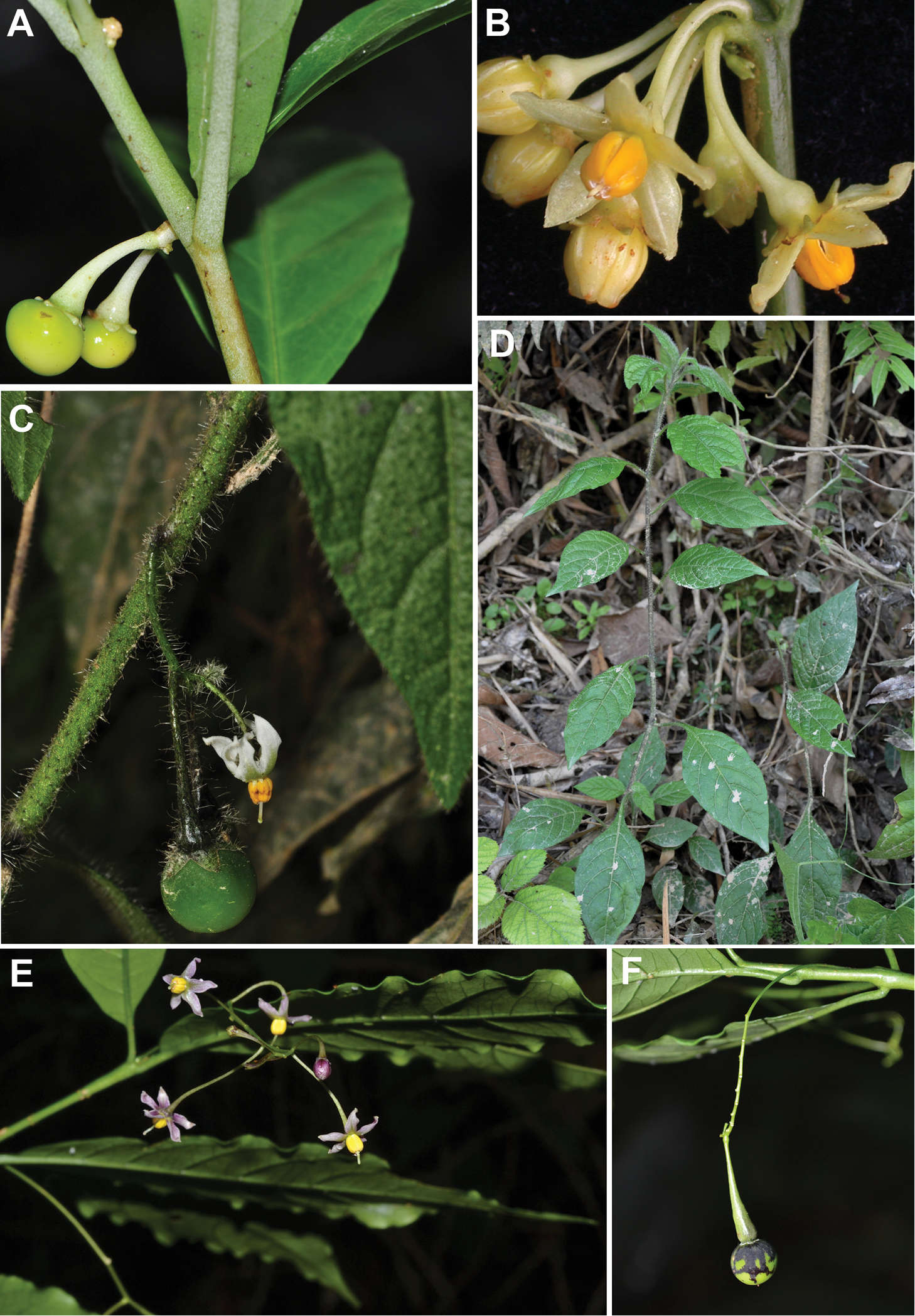
Figure 1.Photograph of living plants of Solanum amorimii, Solanum apiahyense and Solanum filirhachis. A Immature fruit of Solanum amorimii (Giacomin et al.1962) B Flowers of Solanum amorimii (Amorim et al. 5210) C Inflorescence with flower and fruit of Solanum apiahyense (Giacomin et al. 1086) D Habit of Solanum apiahyense (Giacomin et al. 1086) E Inflorescence, flower and leaves of Solanum filirhachis (Giacomin et al. 1854) F Fruit (immature) of Solanum filirhachis (Giacomin et al. 1854). Photographs: A (S. Knapp), B (A.M. Amorim), C–F (L.L. Giacomin).
-
Sandra Knapp, João Renato Stehmann, Leandro L. Giacomin
Phytokeys
Figure 4.Lectotype specimen of Solanum apiahyense (Puiggari s. n., WU). Reproduced with permission of the University of Vienna.