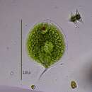
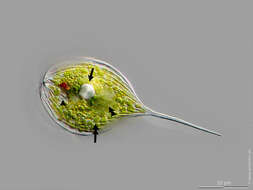
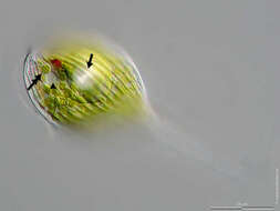
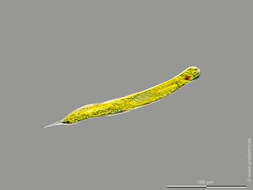
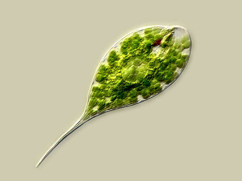
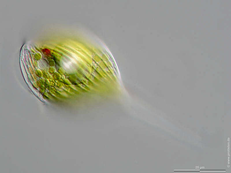
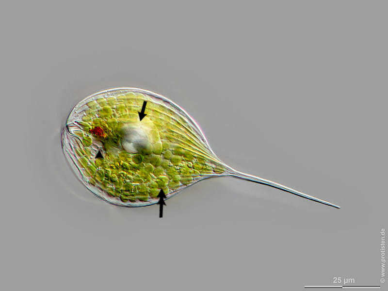
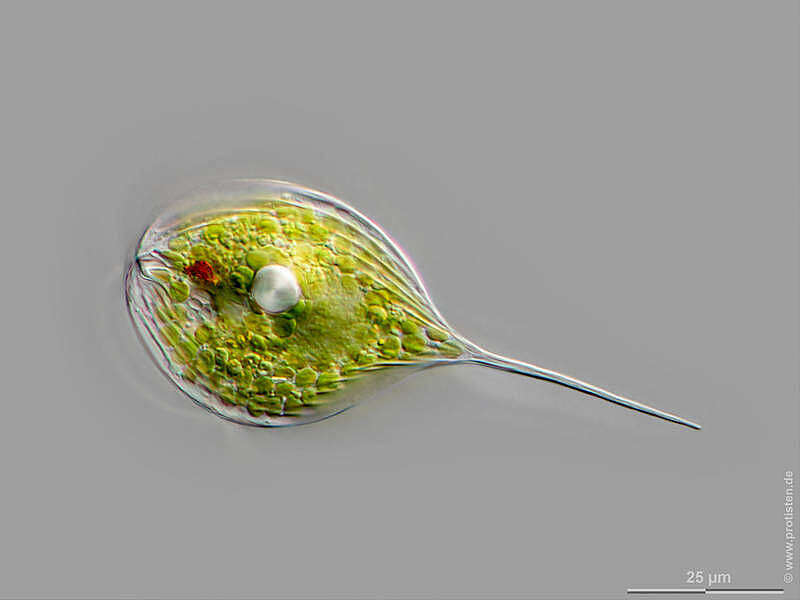
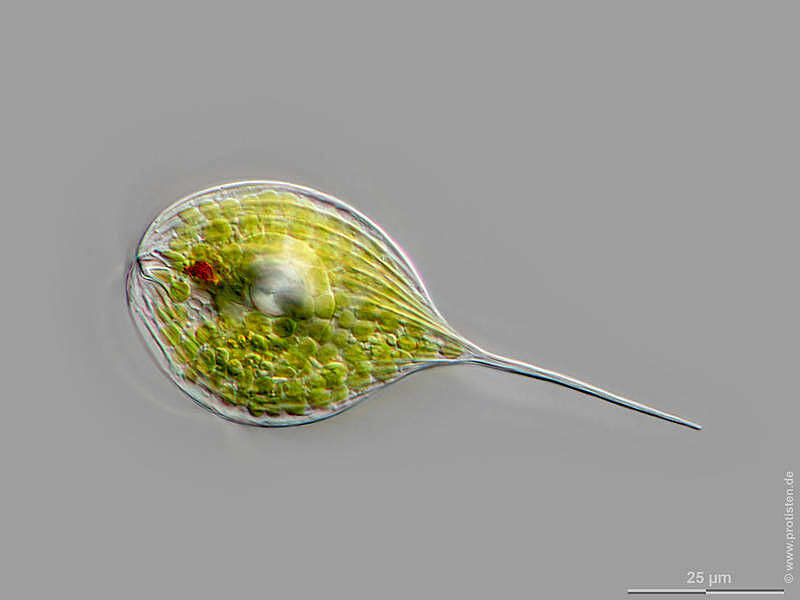
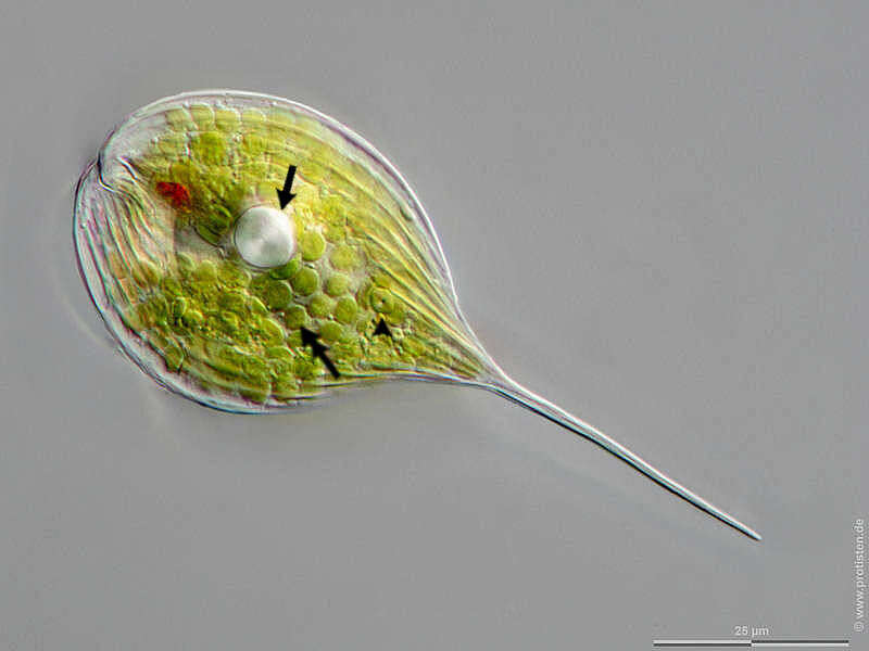
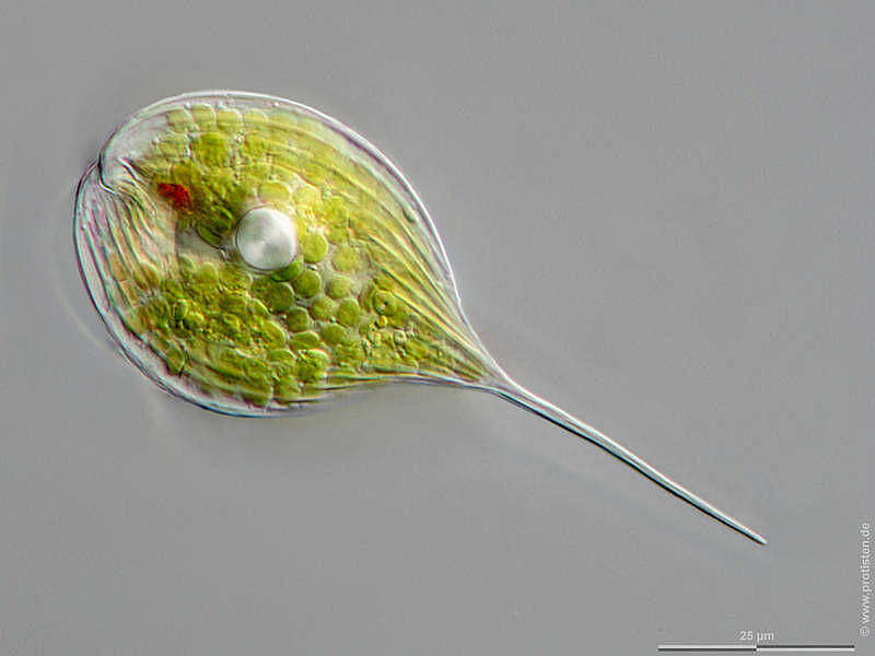
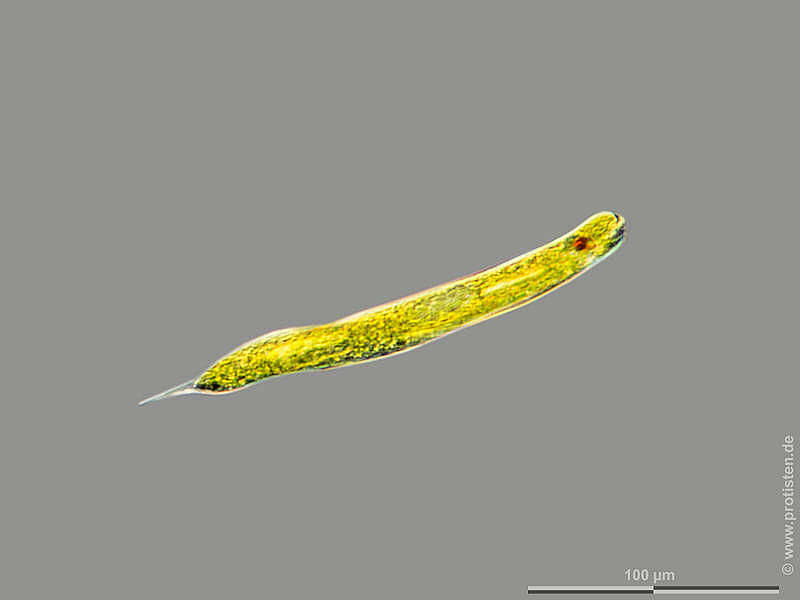
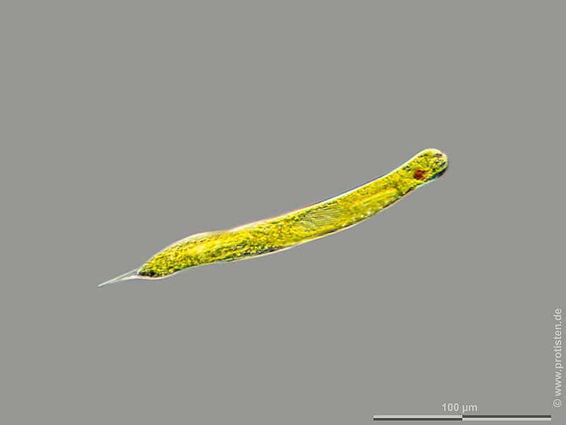
Sampling date 06/2022. Scale bars indicate 25 µm.Two image couples, without and with marking arrows each.First couple:Top view: Red eye spot, paramylon body (arrow), contractile vacuole, fragmented during inflation (arrowhead), disc-shaped chloroplast (double headed arrow).Second couple:Optical cross-section: Red eye spot, paramylon body (arrow), contractile vacuole, fragmented during inflation (arrowhead), nucleus (double arrowhead), disc-shaped chloroplast (double headed arrow). Please click on < or > on the image edges or on the dots at the bottom edge of the images to browse through the slides!Place name: Pond in the forest of Altenholz-Stift (Schleswig-Holstein, Germany)Latitude: 54.384913 Longitude: 10.125691Microscope Zeiss Axioplan, camera Olympus OM-D M5 MKII. DOF images.© Wolfgang Bettighofer,images under Creative Commons License V 3.0 (CC BY-NC-SA).For permission to use of (high resolution) images please contact
postmaster@protisten.de.For further information about the image, please click here:
Link to protisten.de page