-
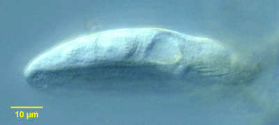
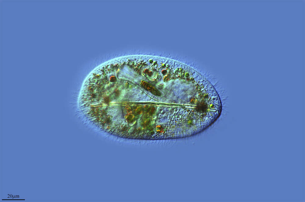
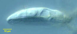
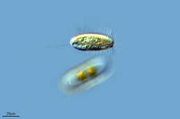
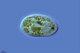
Gastronauta membranaceus (Engelmann in Butschli, 1889). Seen from the side, the body is dorso-ventrally flattened.
-
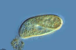
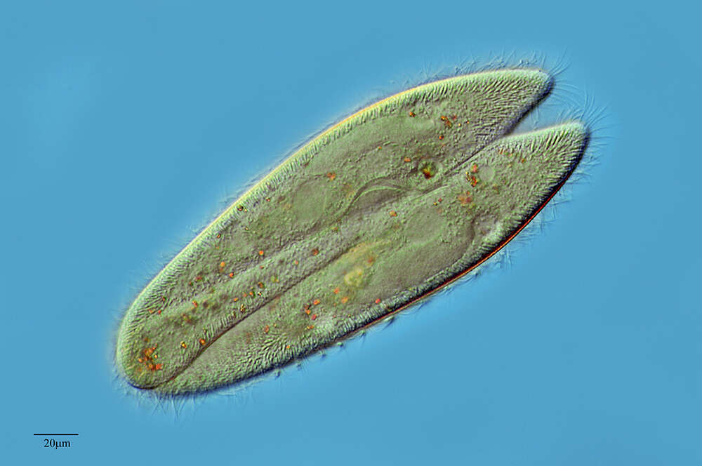
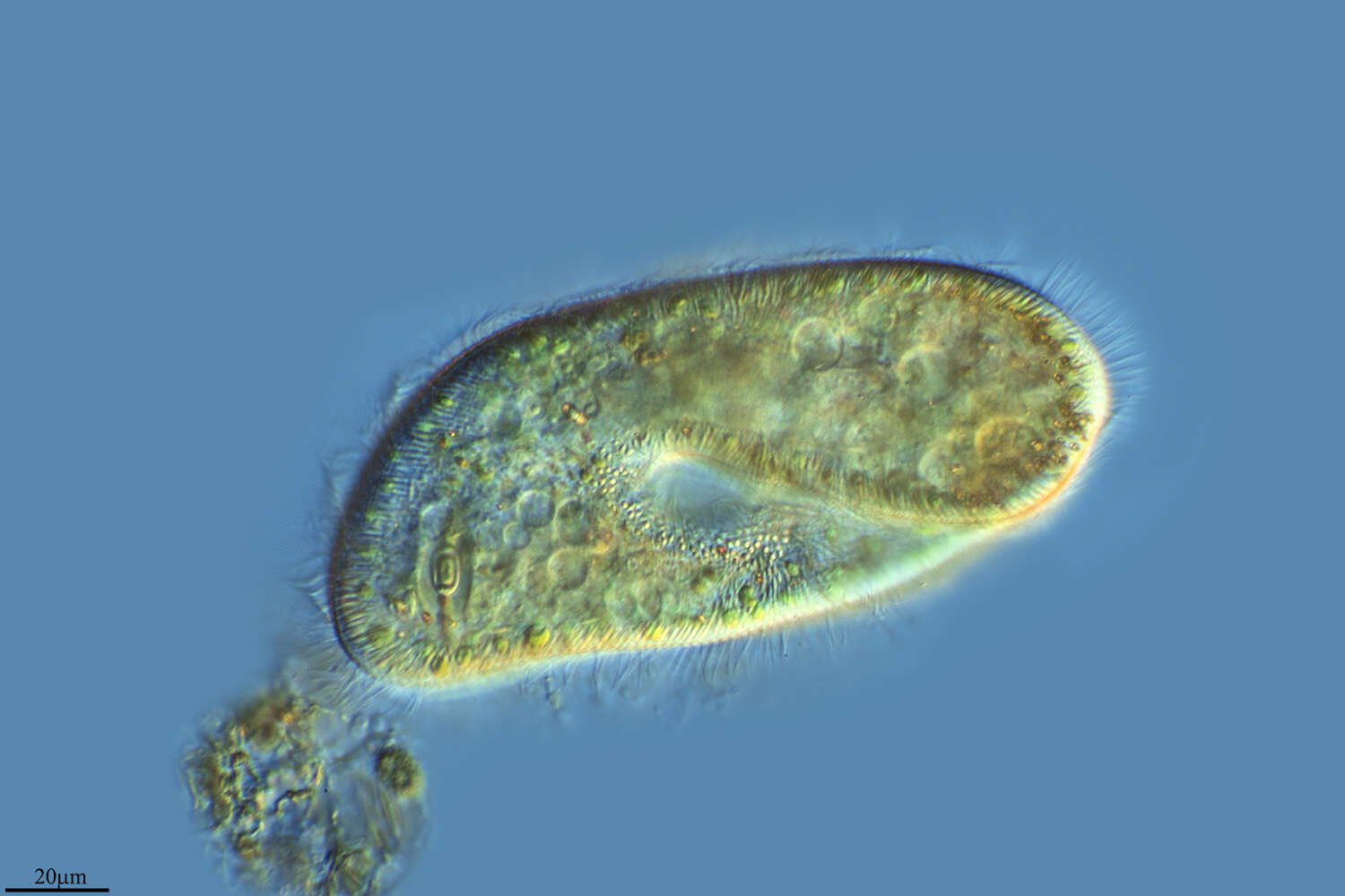
Ventral infraciliature of the hymenostome ciliate Colpidium kleini (Foissner, 1969). C. kleini is very similar in overall appearance to C. colpoda although usually more slender and with fewer somatic kineties. The cytostome is in the anterior 1/4 of the cell. There is a curved paraoral membrane along the convex right margin of the cytostome. The left margin is slightly concave. There are three adoral membranelles. There are 32 to 44 somatic kineties. The kineties to the right and left of the oral aperture meet at a curved preoral suture. The right somatic kineties bend leftward at the level of the cytostome.There is an anterior apical area bare of cilia. There are rows of inconspicuous mucocysts between the somatic kineties. The ellipsoid macronucleus and adjacent micronucleus are centrally located. The single contractile vacuole is located in the midbody with a single excretory pore on the right surface. The feature most clearly distinguishing Colpidium kleini from C. coploda is the silverline system (as demonstrated by silver nitrate staining).Stained by the silver carbonate technic (see Foissner, W.Europ. J. Protistol.27,313-330;1991). Collected from an organically enriched freshwater pond near Boise, Idaho. Brightfield.
-
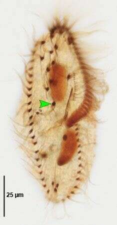
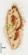
Gonostomum affine (Stein, 1859) Sterki, 1878. Ventral infraciliature. Green arrowhead marks posterior-most "postoral" cirrus. Non-flooded Petri dish soil sample collected from flood-irrigated lawn in Boise, Idaho, June 2008. Protargol (Wilbert modification). Brightfield.
-
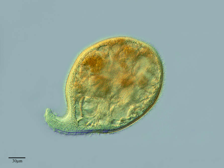
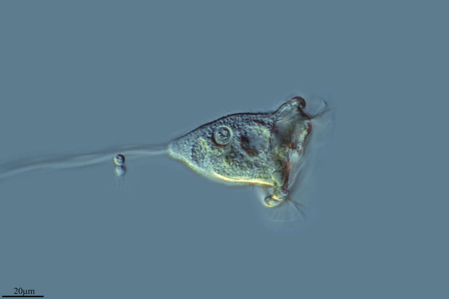
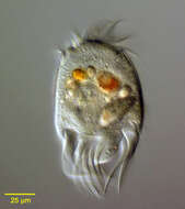
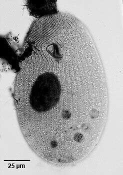
Portrait of the marine hypotrich ciliate, Uronychia transfuga (Müller,1786). Cell outline is ovoid with a colorless rigid pellicle.The broad peristome (not well seen here) is bordered on the left by an adoral zone of membranelles. There are three posterior concavities. Three massive caudal cirri arise from the rightmost concavity. Five transverse cirri arise from the central concavity and three marginal cirri arise from the left concavity (two large and one small). The moniliform macronucleus is arranged in a C-shape.Slow swimming is interrupted by sudden jumping movement. several colorful food vacuoles are visible here. Collected from a commercial saltwater aquarium in Boise, Idaho February 2004. DIC .
-
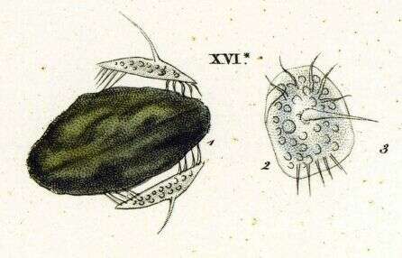
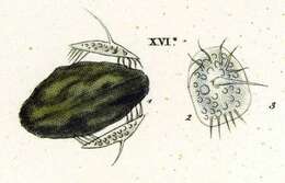
Originally described by Ehrenberg under the name Euplotes turritus.
-
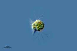
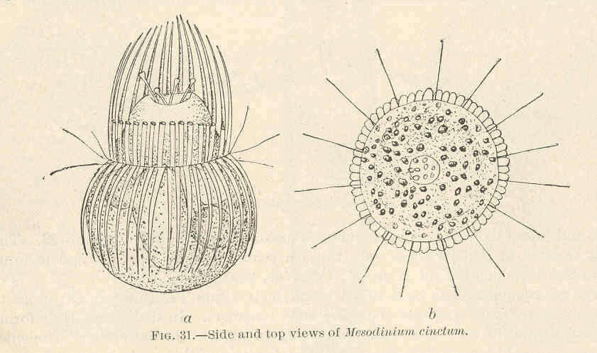
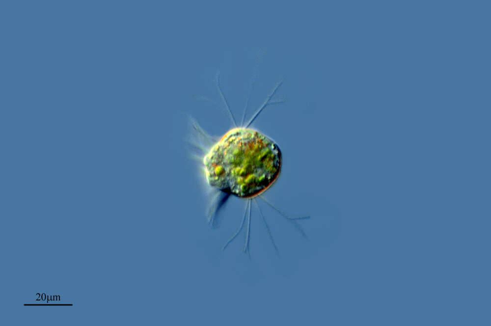
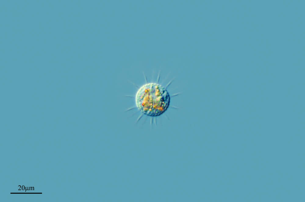
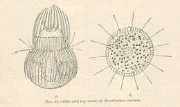
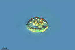
Mesodinium cinctum--Side and top views.
-
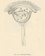
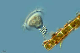
Vorticella patellina.
-
-
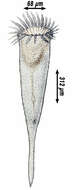
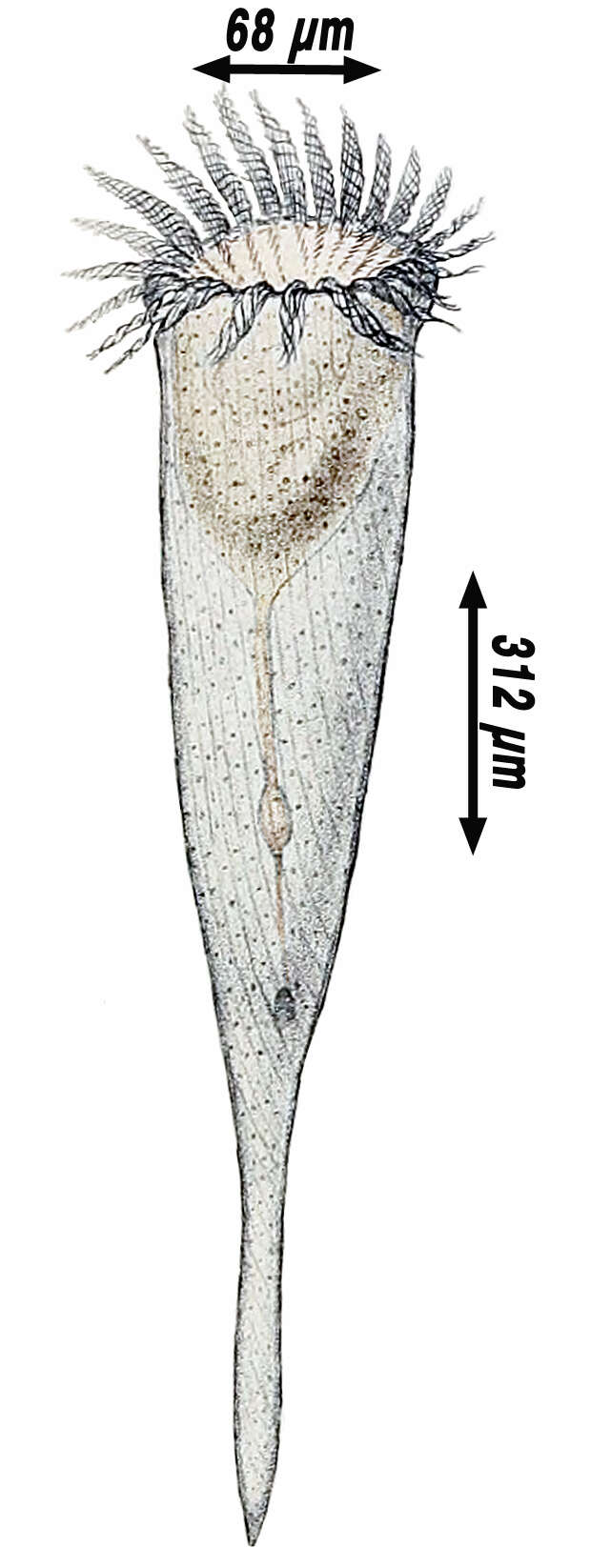
Hermann Fol's drawing from the original description (1881) with his corrected dimensions (1884). False brown color added to the ciliate cell
-
Hoyo de Manzanares, Madrid, Spain
-
Urbanitzacio El Lledoner, Catalonia, Spain
-
Peniscola, Valencia, Spain
-
Castille and Leon, Spain
-
Mohedas de la Jara, Castille la Mancha, Spain
-
A Veiga, Galicia, Spain
-
Lardero, La Rioja, Spain
-
Mahide, Castille and Leon, Spain
-
Ribadelago de Franco, Castille and Leon, Spain
-
Melgar de Tera, Castille and Leon, Spain
-
Villoslada de Cameros, La Rioja, Spain
-
Vitoria, Basque Country, Spain
-
Ribadelago de Franco, Castilla y Len, Espaa
-
Logrono, La Rioja, Spain
-
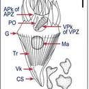
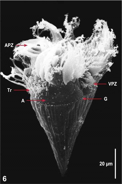
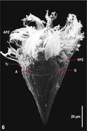
Fig 6 SEM of Lugol?s fixed cell, right lateral view, shows the extrusion of trichites (Tr) and the array (A), where many trichites are located below the surface.