-
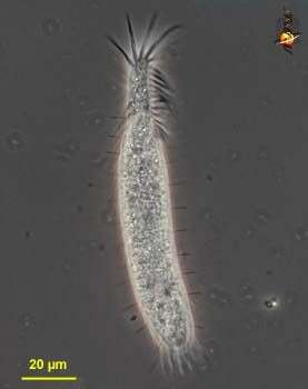
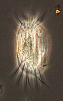
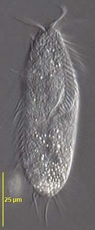
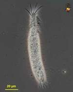
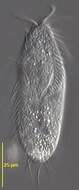
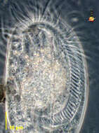
Trachelostyla (track-ell-owe-style-a), a hypotrich ciliate. With a narrowed anterior end to the body, with an adoral zone of membranelles. Phase contrast micrograph.
-
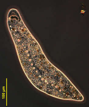
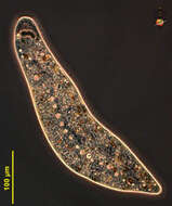
Homalozoon, a elongate ribbon-like predatory ciliate. The body is truncated (cut) off at the front end where the mouth is located, and pointed posteriorly. It has rows of cilia mostly on the ventral side, it glides over the substrate, sometimes contracting. Feeds on detritus and other protists. This image shows the line of contractile vacuoles which extends along the body. Flattened. Phase contrast micrograph.
-
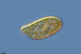
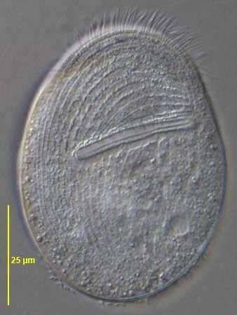
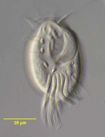
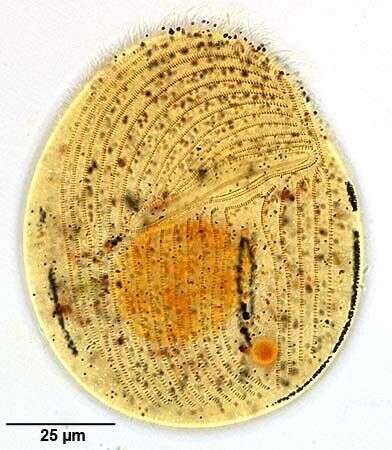
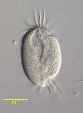
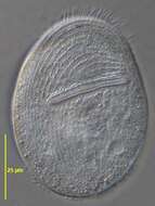
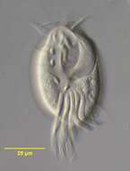
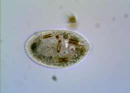
Ventral view (in vivo) of Gastronauta derouxi (FOISSNER & BLATTERER, 1992). Collected from an ephemeral puddle on a grass lawn in Boise, Idaho. July 2007.DIC.
-
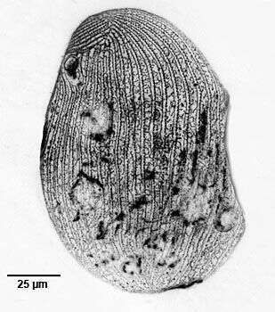
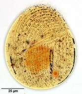
Left lateral view of the silverline system of the hymenostome ciliate Colpidium kleini (Foissner, 1969). C. kleini is very similar in overall appearance to C. colpoda although usually more slender and with fewer somatic kineties. The cytostome is in the anterior 1/4 of the cell. There is a curved paraoral membrane along the convex right margin of the cytostome. The left margin is slightly concave. There are three adoral membranelles. There are 32 to 44 somatic kineties. The kineties to the right and left of the oral aperture meet at a curved preoral suture. There is an anterior apical area bare of cilia. There are rows of inconspicuous mucocysts between the somatic kineties. The ellipsoid macronucleus and adjacent micronucleus are centrally located. The single contractile vacuole is located in the midbody with a single excretory pore on the right surface. The feature most clearly distinguishing Colpidium kleini from C. coploda is the silverline system. In C. kleini there is only one secondary meridian (silverline) between two primary meridians (primary meridians correspond to somatic kineties seen here as the wavier lines). Short transverse L or T-shaped branches arise from both primary and secondary meridians at irregular intervals. In some cases short segments of the secondary meridians may be duplicated. Stained by the dry silver nitrate technic (see Foissner, W.Europ. J. Protistol.27,313-330;1991). Collected from an organically enriched freshwater pond near Boise, Idaho. Brightfield. Black and white.
-
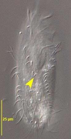
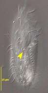
Ventral infraciliature of the oxytrichid,Gonostomum strenuum (ENGELMANN, 1862) STERKI, 1878. G. strenuum differs from G. affine by the greater number of frontoterminal and frontoventral cirri. In G. strenuum the last frontoventral cirrus is just posterior to the peristome (yellow arrowhead).Collected from a non-flooded Petri dish culture of soil from a park lawn in Boise, Idaho. January 2007.DIC.
-
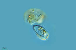
A species of the marine hypotrich genus, Diophrys (DUJARDIN,1840). The genus contains many species. Collected from a commercial marine aquarium in Boise, Idaho. DIC.
-
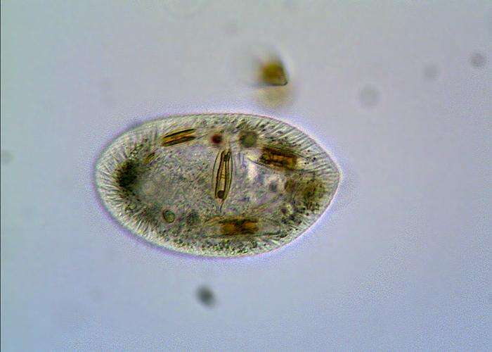
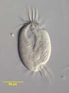
Euplotes (you-p-low-tees) is a hypotrich ciliate. The hypotrichs form part of the spirotrichs, and most have a large adoral zone of membranelles curving around the front of the cells and terminating at the cytostome on the ventral surface. They are called hypotrichs because the cilia that are used for locomotion are located mostly on the ventral side of the cilia. The cilia are clustered into aggregates called cirri. They are clearly evident in this image of the dorsal side of the cell. Euplotids feed on suspended particles such as bacteria and algae. Common. Phase contrast.
-
-
Pera, Faro, Portugal
-
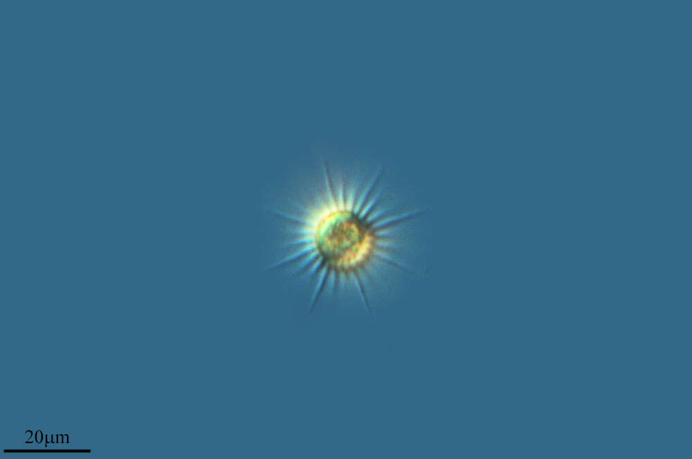
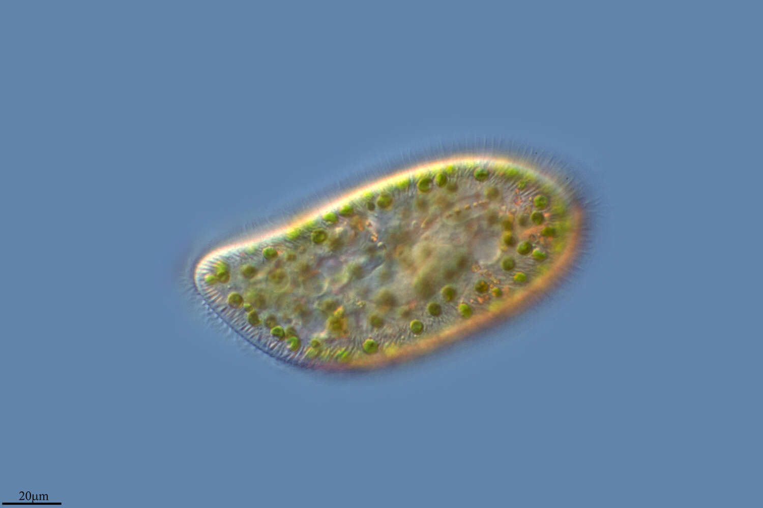
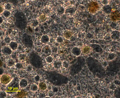
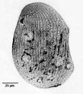
Ovoid, 60-100 micron long. Numerous large trichocysts.
-
Villoslada de Cameros, La Rioja, Spain
-
Ribadelago, Castille and Leon, Spain
-
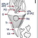
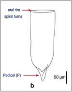
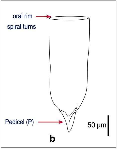
Fig1b: Favella ehrenbergii Line drawing of lorica morphology after Kofoid & Campbell, 1929
-
-
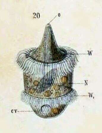
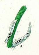
Originally described by Schewiakoff under the name Didinium balbianii. Schewiakoff describes this as an individual apprehended in division. o--Mouth N--Macronucleus W--Ciliated ring W1--New ciliated ring, prepared for offspring
-
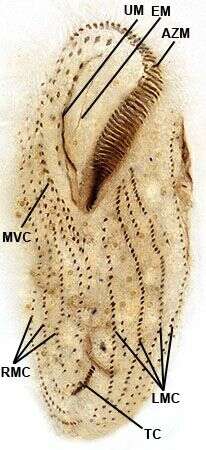
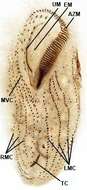
Ventral infraciliature of the large hypotrich ciliate, Urostyla grandis (EHRENBERG,1830). A file of buccal cirri parallels the undulating membrane (UM).Em=endoral membrane.AZM=adoral zone of membranelles.There is a zig-zag file of midventral cirri (MVC) between the right and left marginal cirral rows. There is an obliquely oriented row of about 12 transverse cirri (TC).Collected from a freshwater canal near Boise, Idaho.Stained by the protargol A technique (see Foissner, W. Europ. J. Protistol., 27:313-330;1991).Brightfield.
-
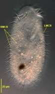
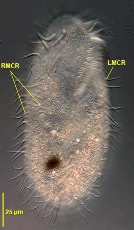
Ventral view of Pleurotricha lanceolata (EHRENBERG,1835) STEIN, 1859. RMCR= two right marginal cirral rows. LMCR=single left marginal cirral row.Collected from a flood-irrigated grass lawn in Boise,Idaho. May 2008. DIC.
-
Homalozoon, a elongate-ribbon like predatory ciliate. The body is truncated (cut) off at the front end where the mouth is located, and pointed posteriorly. It has rows of cilia mostly on the ventral side, it glides over the substrate, sometimes contracting. Feeds on detritus and other protists. This image shows the macronucleus, which takes to form of a row of ellip[tical beads attached end-to-end. The micronuclei are small round dark structures adjacent to the macronucleus. Phase contrast micrograph.
-
Ventral infraciliature of Gastronauta derouxi (FOISSNER & BLATTERER, 1992).Collected from an ephemeral puddle on a grass lawn in Boise, Idaho. July 2007. Stained by the silver carbonate technique (Foissner,W. Europ. J. Protistol.27:313-330;1991).Brightfield.
-
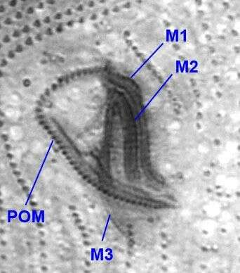
Oral infraciliature of Colpidium kleini (FOISSNER, 1969).There are three adoral membranelles (M1-3) and a right paraoral membrane (POM).Stained by the silver carbonate technique (see Foissner, W. Europ. J. Protistol., 27:313-330;1991).Brightfield.
-
Ventral view of the oxytrichid,Gonostomum strenuum (ENGELMANN, 1862) STERKI, 1878. G. strenuum differs from G. affine by the greater number of frontoterminal and frontoventral cirri. In G. strenuum the last frontoventral cirrus is just posterior to the peristome .Collected from a non-flooded Petri dish culture of soil from a park lawn in Boise, Idaho. January 2007.DIC.
-
A species of the marine hypotrich genus, Diophrys (DUJARDIN,1840). The genus contains many species. Collected from a commercial marine aquarium in Boise, Idaho. DIC.
-
Euplotes (you-p-low-tees) is a hypotrich ciliate. The hypotrichs form part of the spirotrichs, and most have a large adoral zone of membranelles curving around the front of the cells and terminating at the cytostome on the ventral surface. They are called hypotrichs because the cilia that are used for locomotion are located mostly on the ventral side of the cilia. The cilia are clustered into aggregates called cirri. This image illustrates the adoral zone of membranelles. Phase contrast.
-