-
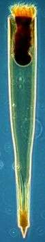
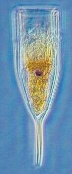
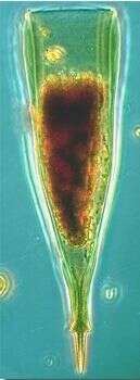
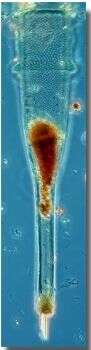
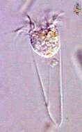
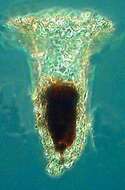
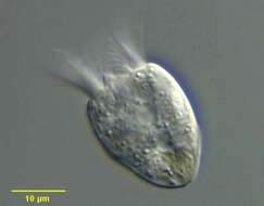
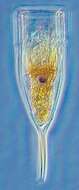
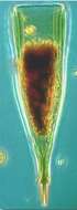
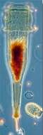
Eutintinnus (you-tin-tin-us), one of the tintinnid ciliates. These are mostly marine choreotrichs in which the cell is located within an open lorica which it drags around while it swims. The different genera and species are mostly distinguished by the different appearances of the lorica. This genus has a conical lorica which is open at both ends. With an adoral zone of membranelles located around the top end of the cell. Differential interference contrast.
-
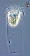
Eutintinnus (you-tin-tin-us), one of the tintinnid ciliates. These are mostly marine choreotrichs in which the cell is located within an open lorica which it drags around while it swims. The different genera and species are mostly distinguished by the different appearances of the lorica. This genus has a conical lorica which is open at both ends. With an adoral zone of membranelles located around the top end of the cell. Phase contrast.
-
-
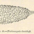
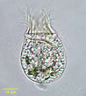
Tintinnopsis campanula.
-
-
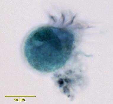
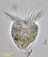
Strobilidium caudatum (Fromental, 1874) Foissner, 1987, a spirotrich ciliate. Synonym of S. gyrans.The cell body is goblet shaped with a complete circular (closed type) wreath of membranelles at the anterior end. The adoral zone of membranelles is well seen in this image. Unlike Strombidium, there is no posterior lorica. There are five very reduced rows of somatic cilia which converge at the posterior end of the cell to form a tight spiral as seen in this image. The terminus of the cell secretes a mucus thread allowing attachment to the substrate. Once attached the cell moves back and forth in pendulum fashion for a time then breaking free to swim away very rapidly. The oral aperture is eccentrically located within the adoral zone of membranelles. A peripheral contractile vacuole is seen posterolaterally in this image. Feeds on diatoms, flagellates and probably bacteria. From freshwater pond near Boise, Idaho. Brightfield illumination.
-
Anterior apical view of the adoral zone of membranelles of Strobilidium caudatum (Fromental,1874) Foissner,1987, a spirotrich ciliate. Synonym of S. gyrans.Stained by the silver carbonate technique (see Foissner, W. Europ. J. Protistol., 27:313-330;1991).Brightfield.
-
Strobilidium caudatum (FROMENTAL, 1874) FOISSNER, 1987, a spirotrich ciliate. Synonym of S. gyrans.The cell body is goblet shaped with a complete circular (closed type) wreath of membranelles at the anterior end. The adoral zone of membranelles is well seen in this image. Unlike Strombidium, there is no posterior lorica. There are five very reduced rows of somatic cilia which converge at the posterior end of the cell to form a tight spiral as seen in this image.Three of these are seen in this image (red arrowheads).The posterior terminus of the cell secretes a mucus thread allowing attachment to the substrate. Once attached the cell moves back and forth in pendulum fashion for a time then breaks free to swim away very rapidly. The cytostome is eccentrically located within the adoral zone of membranelles. From freshwater pond near Boise, Idaho. Protargol (see Foissner, W. Europ. J. Protistol., 27:313-330;1991).Brightfield.
-

Detail of somatic ciliary row of the oligotrich ciliate, Rimostrombidium hyalinum. This genus is distinguished from the similar Strombidium by the small size and the termination of the spiral somatic ciliary rows before they reach the posterior pole of the cell (in Strombidium they form a terminal spiral). R. hyalinum is colorless. The cell body is transversely truncate anteriorly and bluntly conical posteriorly. The cytostome is at the anterior end. A circular adoral zone of membranelles is present (seen well here). There are six indistinct, slightly spiraling rows of short somatic cilia that extend nearly the length of the body. It is seldom possible to keep more than one row in the focal plane in the lateral view (part of one row is seen here on viewer's left). The C- shaped macronucleus is anterior and transversely oriented (not seen here). The peripheral contractile vacuole is located in the posterior of the cell (not seen here). Collected from freshwater pond near Boise, Idaho September 2003. DIC optics.
-
Methyl green-Pyronin stained preparation of the oligotrich ciliate, Rimostrombidium hyalinum demonstrating the C-shaped transversely oriented anterior macronucleus. The small spherical micronucleus is superimposed on the center of the macronucleus in this image. Collected from freshwater pond near Boise, Idaho September 2003. DIC optics.
-
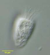
Portrait of the oligotrich ciliate, Romstrombidium hyalinum. This genus is distinguished from the similar Strombidium by the small size and the termination of the spiral somatic ciliary rows before they reach the posterior pole of the cell (in Strombidium they form a terminal spiral). There are several species. R. hyalinum is colorless. The cell body is transversely truncate anteriorly and bluntly conical posteriorly. The cytostome is at the anterior end. A circular adoral zone of membranelles is present. There are six indistinct, slightly spiraling rows of short somatic cilia that extend nearly the length of the body. It is seldom possible to keep more than one row in the focal plane in the lateral view. The C- shaped macronucleus is anterior and transversely oriented The peripheral contractile vacuole is located in the posterior of the cell. A food vacuole is seen here posteriorly. Collected from freshwater pond near Boise, Idaho September 2003. DIC optics.
-
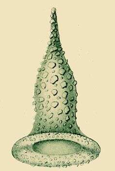
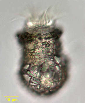
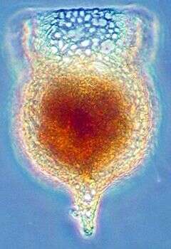
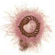
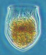
Codonella cratera lorica. Codonella cratera is a large tintinnid ciliate. A distinctive chitinous lorica is coated with xenosomes (foreign particles such as sand particles as in these individuals and diatoms). The posterior lorica is broadly spherical with a cylindrical anterior half. There is a prominent circumferential anterior adoral zone of membranelles. From freshwater pond near Boise, Idaho. Oblique illumination.
-
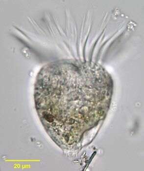
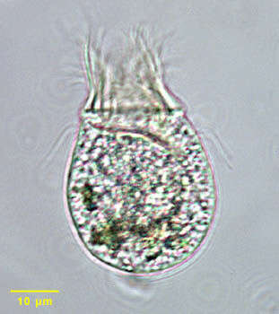
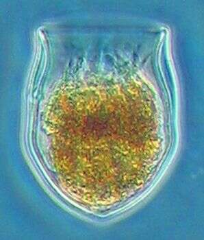
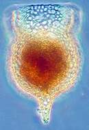
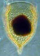
Codonella cratera (Leidy, 1877) Imhof, 1885, which has fled its lorica. The cell body is trumpet shaped when in its lorica, attaching to the lorica base by a drawn-out posterior extension. When free-swimming, the cell body assumes a more globular shape. There is a prominent circumferential anterior adoral zone of membranelles. The buccal cavity is funnel-shaped. Somatic ciliature is uniform and slightly spirals from anterior to posterior. The macronucleus is bipartite. There is a single anterior contractile vacuole. Codonella appears to be omnivorous. Ingested algae are visible in this image. From freshwater pond near Boise, Idaho. Brightfield illumination.
-
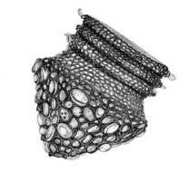
This ciliat belonging to the morphological group of oligotrichids builds marvellous amphora shaped loricae putting together mostly centric diatom frustules. This drawing (it´s an original) was inspired by a SEM picture of a good friend. Collection from littoral region (stand of Phragmites) of oligotrophic lake near Kiel (Schleswig-Holstein, Germany).
-
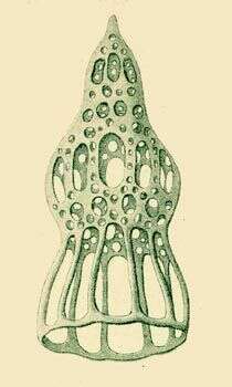
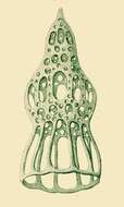
Dictyocysta tiara.
-
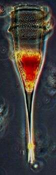
This tintinnid is about 400 microns long. This Lugol's-preserved specimen was found in samples from the South Pacific
-
Xystonellopsis dicymatica is a tropical tintiinid ciliate about 350 microns long. The image is of a lugol's preserved specimen ffrom the South East Pacific.
-
-
-
-
-
-
-