-
-
-
-
-
-
-
-
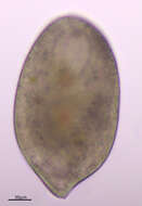
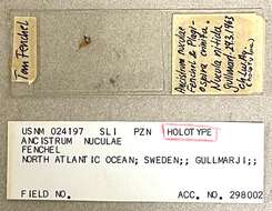
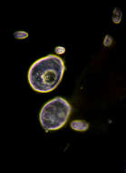
USNM POR 24197 slide image
-
-
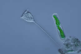
[taxonomy:genus=Vorticella]
Date:
23 Aug 2011
Location:
Small lake in Kent Ridge Park. Water margin with vegetation, brown sediment with organic detritus. Tadpoles resting nearby.
Microscope:
Bright-field with closed condenser aperture.
Camera:
Nikon D7000
Collector:
Brandon Seah
Scale:
20830 pixels/mm = 20.8 pixels/µm
-
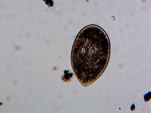
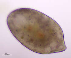
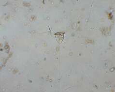
[taxonomy:genus=Disematostoma]
Date:
7 Sep 2011, originally collected 6 Sep 2011
Location:
Freshwater stream flowing out of MacRitchie Reservoir, close to Venus Drive entrance. On field trip with NUS freshwater biology class. Stream was brown with sandy bottom, water mostly clear.
Microscope:
Bright-field with closed condenser aperture.
Camera:
Nikon D7000
Collector:
Brandon Seah
Scale:
20830 pixels/mm = 20.8 pixels/µm (40x)
-
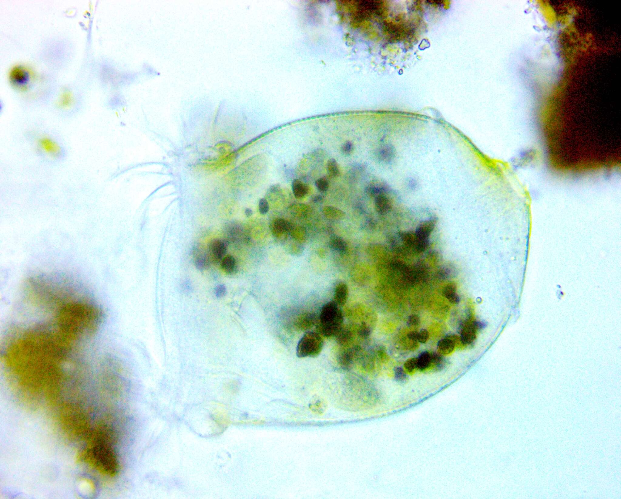
[taxonomy:genus=Cinetochilum]
A scuticociliate with ventral ciliature and a flattened-oval body. It swims around and also uses its cilia to "walk" on substrates, as this video shows. Three long "tail" (caudal) cilia.
-
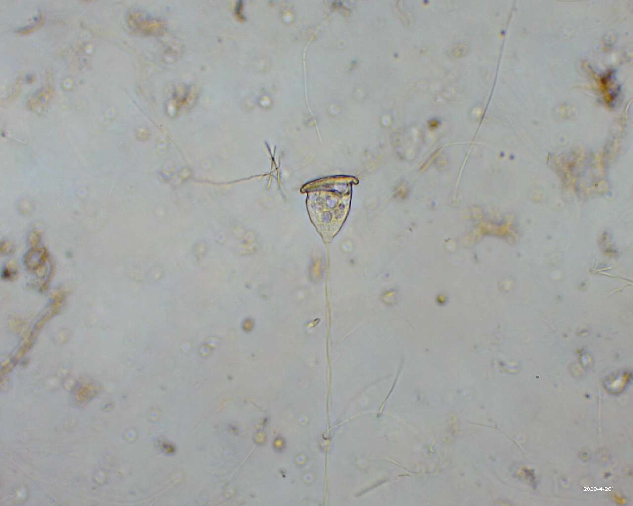
[taxonomy:genus=Thuricola]
This is a ciliate living inside a vase-shaped "lorica", or shell, that comes with its own lid that shuts behind the cell when it retracts into its den. Unfortunately, my attempt to "tap on the glass" and have it retract were not successful. Note the streaming of particles in the water due to the feeding current set up by the cilia of the creature.
-
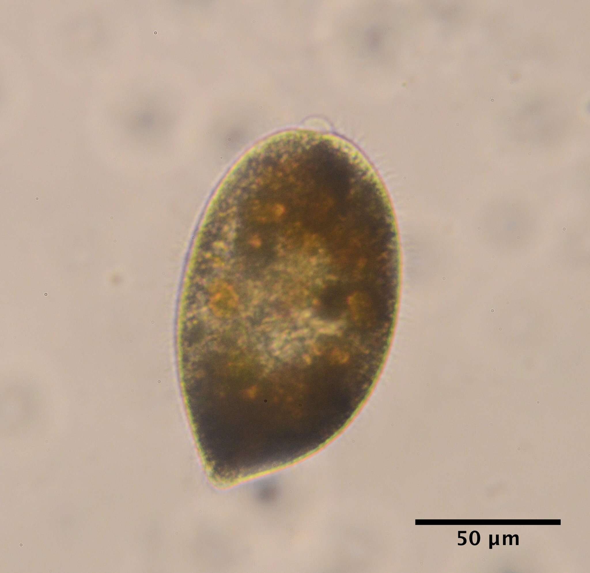
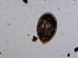
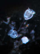
[taxonomy:genus=Urocentrum]
Date:
7 Sep 2011, originally collected 6 Sep 2011
Location:
Freshwater stream flowing out of MacRitchie Reservoir, close to Venus Drive entrance. On field trip with NUS freshwater biology class. Stream was brown with sandy bottom, water mostly clear.
Microscope:
Bright-field with closed condenser aperture.
Camera:
Nikon D7000
Collector:
Brandon Seah
Scale:
20830 pixels/mm = 20.8 pixels/µm (40x)
-
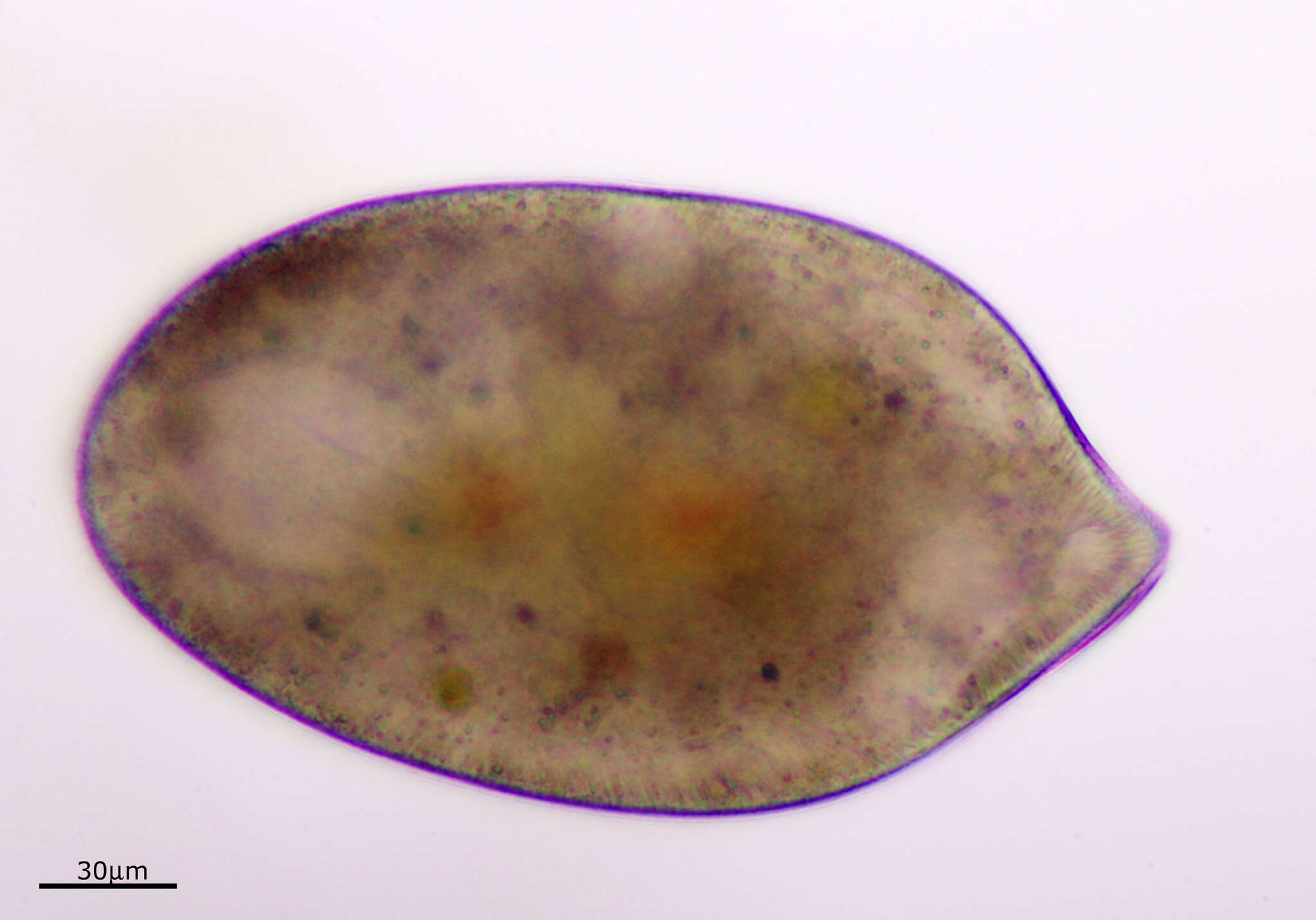
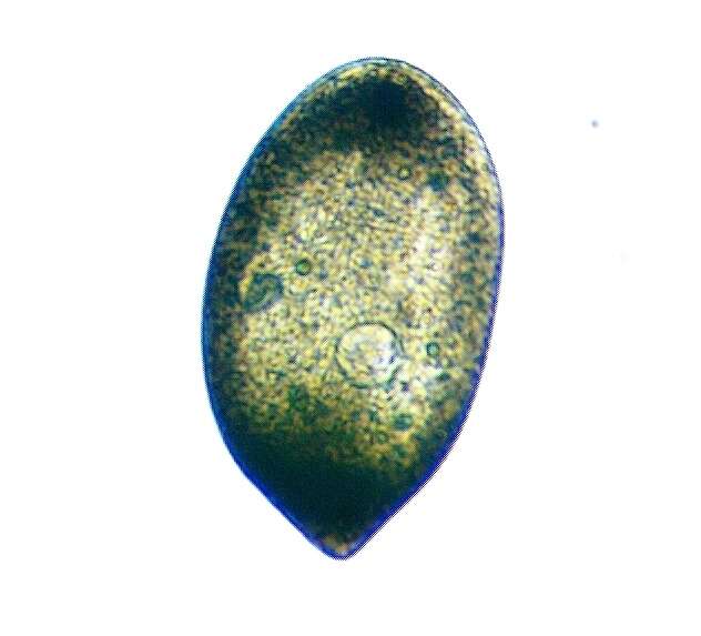
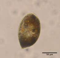
[taxonomy:genus=Cyclidium]
Date:
12 Aug 2011
Location:
Permanently wet longkang beside NUS Enterprise Incubator, under shade of large banyan tree. Drain full of leaf litter and shed banyan roots. Water clear, not green, grey-brown floc when bottom and litter stirred.
Microscope:
Bright-field with closed condenser aperture.
Camera:
Nikon D7000
Collector:
Brandon Seah
-
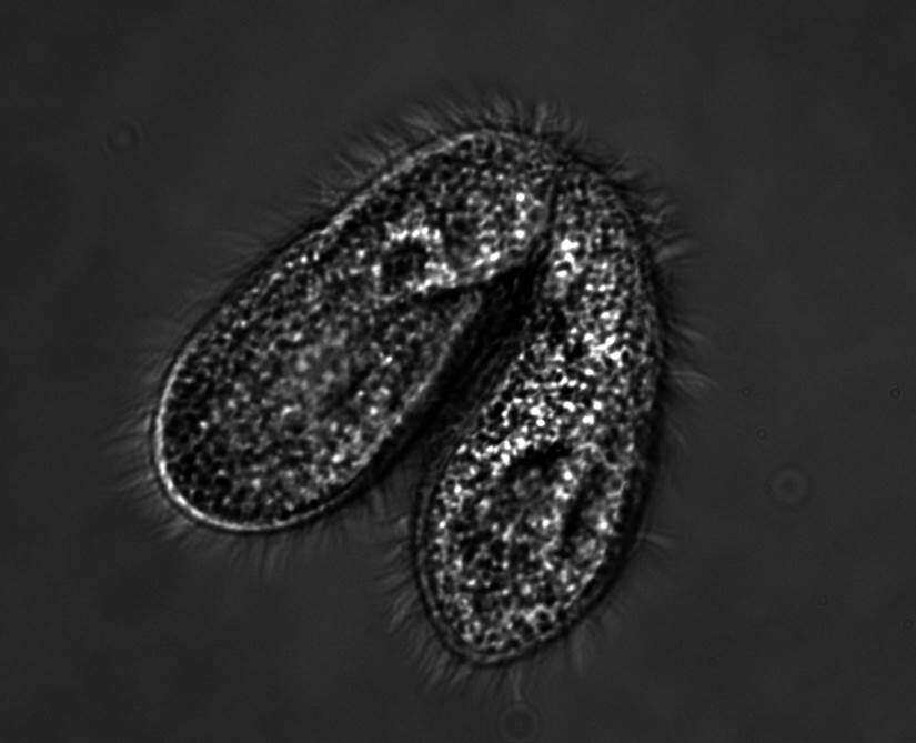
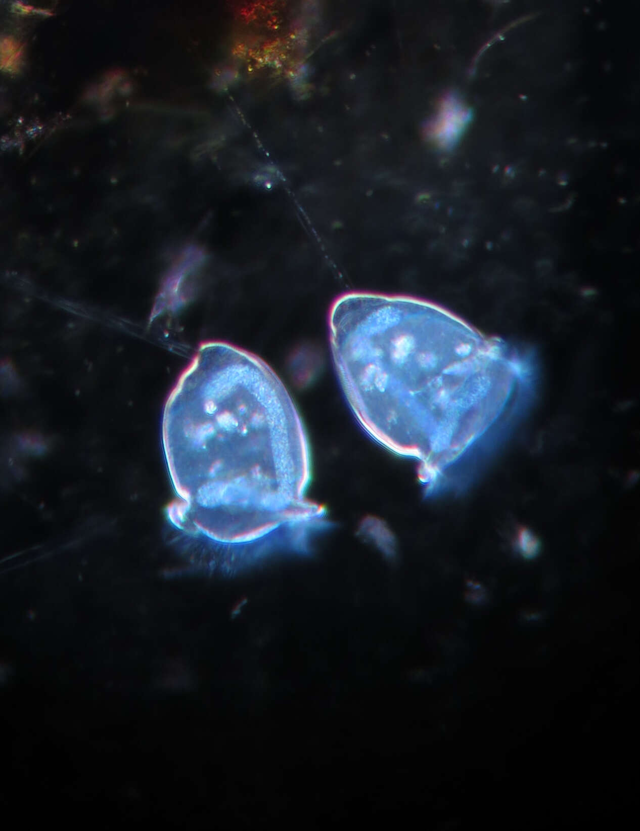
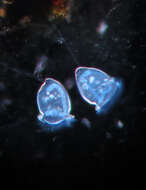
Description: English: Two cells of complementary mating types pair to exchange nuclei during sexual conjugation. Date: 8 March 2016. Source: Own work. Author:
Jmf368w.
-
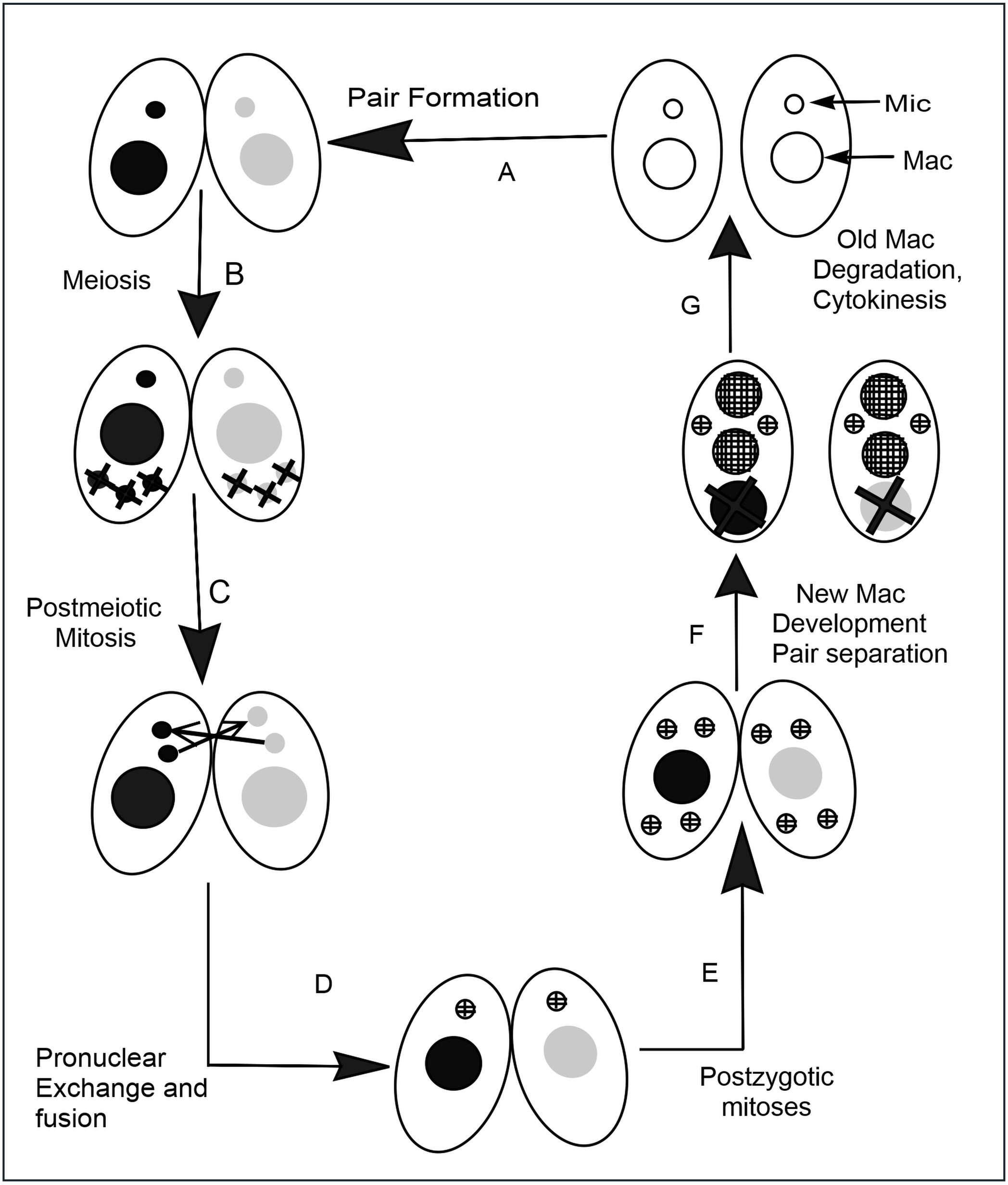
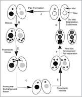
Description: English: Tetrahymena conjugation When nutrients are scarce, two individuals (A) pair with each other and begin sexual reproduction (conjugation). (B) The diploid micronucleus in each individual undergoes meiosis to form four haploid nuclei, and three of these are degraded. (C) The remaining haploid nucleus divides mitotically to form two pronuclei in each cell. (D) One of the two pronuclei in each cell is exchanged with the mating partner, and fusion leads to he formation of the diploid zygotic nucleus. (E) The zygotic nucleus divides twice mitotically to form four nuclei. (F) Two nuclei become micronuclei, and the other two differentiate to become macronuclei; the original parental macronucleus is degraded. (G) Cell division occurs and the nuclei are distributed to the daughter cells so that each progeny receives one micronucleus and one macronucleus. Date: 16 April 2014, 11:58:27. Source: Own work. Author:
Chaya5260.
-
-
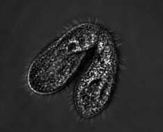
Description: 160x, Dunkelfeld Kernfärbung mit Ethylgrün (Lebendfärbung). Das "J" ist der Zellkern, der bei Protozoen nicht immer die Form einer Kugel hat. Date: 5 November 2012, 11:01. Source:
Glockentierchen. Author:
Picturepest.
-
Summary.mw-parser-output table.commons-file-information-table,.mw-parser-output.fileinfotpl-type-information{border:1px solid #a2a9b1;background-color:#f8f9fa;padding:5px;font-size:95%;border-spacing:2px;box-sizing:border-box;margin:0;width:100%}.mw-parser-output table.commons-file-information-table>tbody>tr,.mw-parser-output.fileinfotpl-type-information>tbody>tr{vertical-align:top}.mw-parser-output table.commons-file-information-table>tbody>tr>td,.mw-parser-output table.commons-file-information-table>tbody>tr>th,.mw-parser-output.fileinfotpl-type-information>tbody>tr>td,.mw-parser-output.fileinfotpl-type-information>tbody>tr>th{padding:4px}.mw-parser-output.fileinfo-paramfield{background:#ccf;text-align:right;padding-right:0.4em;width:15%;font-weight:bold}.mw-parser-output.commons-file-information-table+table.commons-file-information-table,.mw-parser-output.commons-file-information-table+div.commons-file-information-table>table{border-top:0;padding-top:0;margin-top:-8px}@media only screen and (max-width:719px){.mw-parser-output table.commons-file-information-table,.mw-parser-output.commons-file-information-table.fileinfotpl-type-information{border-spacing:0;padding:0;word-break:break-word;width:100%!important}.mw-parser-output.commons-file-information-table>tbody,.mw-parser-output.fileinfotpl-type-information>tbody{display:block}.mw-parser-output.commons-file-information-table>tbody>tr>td,.mw-parser-output.commons-file-information-table>tbody>tr>th,.mw-parser-output.fileinfotpl-type-information>tbody>tr>td,.mw-parser-output.fileinfotpl-type-information>tbody>tr>th{padding:0.2em 0.4em;text-align:left;text-align:start}.mw-parser-output.commons-file-information-table>tbody>tr,.mw-parser-output.fileinfotpl-type-information>tbody>tr{display:flex;flex-direction:column}.mw-parser-output.commons-file-information-table+table.commons-file-information-table,.mw-parser-output.commons-file-information-table+div.commons-file-information-table>table{margin-top:-1px}.mw-parser-output.fileinfo-paramfield{box-sizing:border-box;flex:1 0 100%;width:100%}} Description: Deutsch: lichtmikroskopische Aufnahme eines Glockentierchens. Date: 30 April 2020. Source: Own work. Author:
Imrahil22.
-
Description: 160x, Dunkelfeld Kernfärbung mit Ethylgrün (Lebendfärbung). Das "J" ist der Zellkern, der bei Protozoen nicht immer die Form einer Kugel hat. Date: 5 November 2012, 11:00. Source:
Glockentierchen. Author:
Picturepest.
-


