Jeremy A. Miller, Charles E. Griswold, Nikolaj Scharff, Milan Řezáč, Tamás Szűts, Mohammad Marhabaie
Zookeys
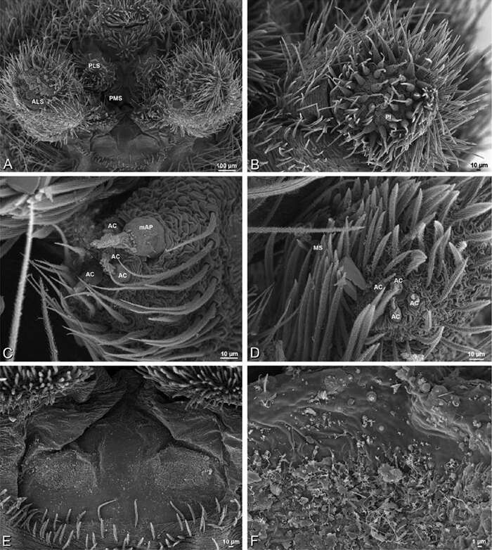
Figure 78.A–F Seothyra henscheli from Gobabeb Station, Namibia (SMN 40828, NMN), scanning electron micrographs of male spinnerets. A overview B right ALS C left PMS D left PLS E vestigial cribellum F detail of vestigial cribellum. AC aciniform gland spigot ALS anterior lateral spinneret mAP minor ampullate gland spigot MS modified spigot PI piriform gland spigot PLS posterior lateral spinneret PMS posterior median spinneret.
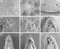
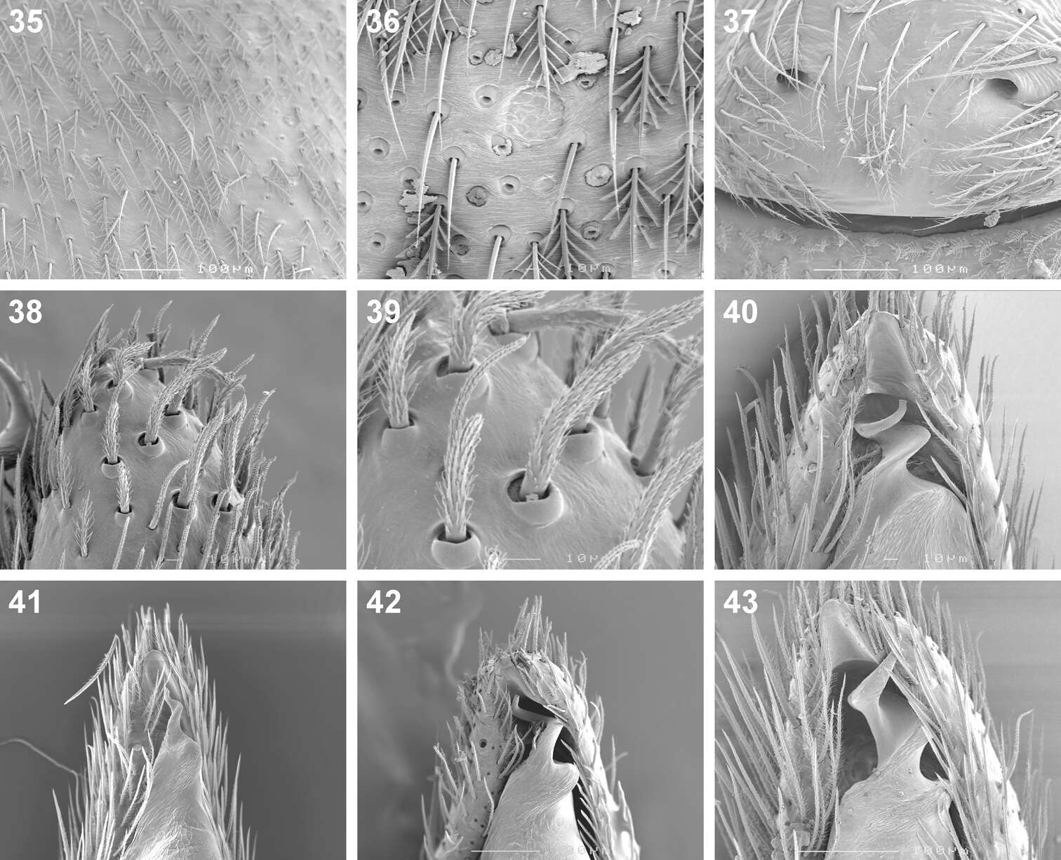
Figures 35–43.Scanning electron microscope photographs of Cambalida dippenaarae sp. n. (35–39, 42), Cambalida compressa sp. n. (40), Cambalida deminuta (Simon, 1909) (41) and Cambalida loricifera (Simon, 1885) (43): 35 female, dorsal abdominal surface 36 dorsal abdominal sigillum and detail of plumose setae 37 female epigyne 38 thickened setae at dorsal distal end of male palpal cymbium 39 detail of modified setae 40–43 male emboli.


