-
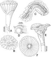
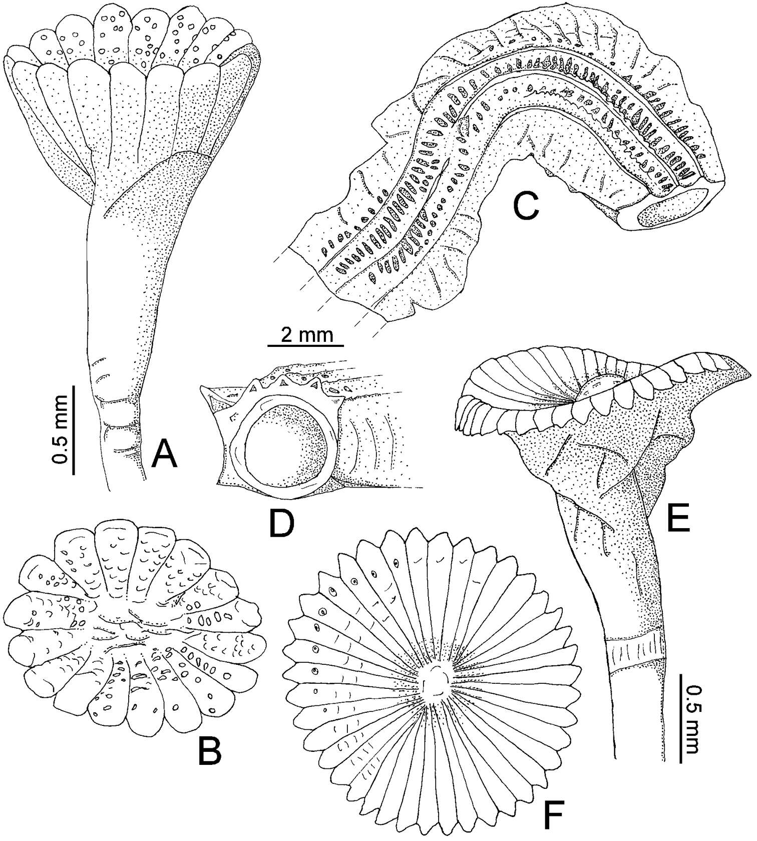
Figure 1.A–D Serpula madrigalae sp. n., from Turks and Caicos Islands, USNM 1157006, holotype A–B operculum in lateral and aboral views C–D tube in dorsal and frontal views E–F Serpula cf. vermicularis, from Nigeria, UMML 22.545 E–F operculum in lateral and aboral views.
-
Ratmanee Chanabun, Chirasak Sutcharit, Piyoros Tongkerd, Somsak Panha
Zookeys
Figure 11.Morphology of the lectotype (ZMH V9301) of Glyphidrilus buttikoferi Michaelsen, 1922, showing the A external ventral and B internal dorsal views.
-
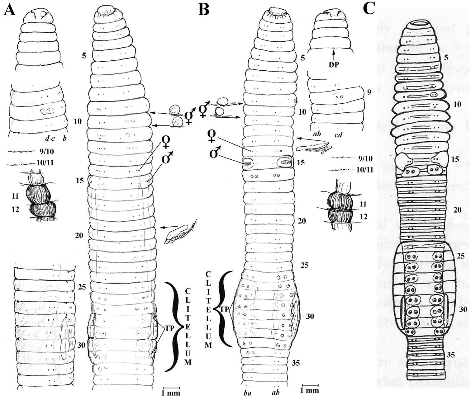
Figure 1.A Eisenia fetida specimen S1 from Jeju Isl., Korea; anterio-ventral and lateral views, dorsal prostomium; spermathecae and calciferous glands in situ, nephridium from 20lhs B Eisenia fetida S3 ditto with nephridium in 13lhs C Athecal Allolobophora hataii Kobayashi, 1940: fig. 5 (incertae sedis) for comparison.
-
Robert J. Blakemore, Seunghan Lee, Wonchoel Lee, Hong-Yul Seo
Zookeys
Figure 1.Amynthas daeari sp. n. showing ventral view with spermathecae, their composite genital marking glands and 18lhs prostate in situ plus incised intestinal caecum in 27; dorsal view of prostomium; [boxed are lateral views of spermathecal pores and male field in 18rhs to same scale].
-
Sergio I. Salazar-Vallejo, Galina Buzhinskaja
Zookeys
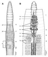
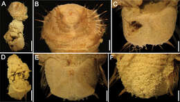
Figure 1.Caulleryaspis fauchaldi sp. n. A Holotype (LACM 5360), ventral view B Anterior end, frontal view C Ventro-caudal shield, frontal view D Paratype (LACM 5361), ventral view E Ventro-caudal shield, frontal view F Posterior end, dorsal view. Bars: A 1.8 mm B, C, E, F 0.6 mm D 2 mm.
-
María E. García-Garza, J.A. de León-González
Zookeys
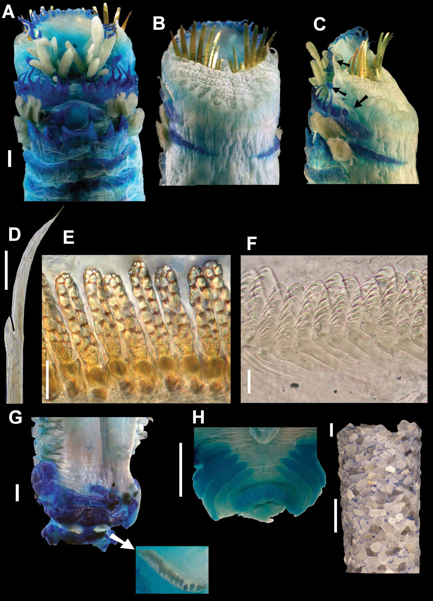
Figure 2.Amphictene helenae. Holotype. A ventral view of anterior end B dorsal view of anterior end C lateral view of anterior end, showing tentacular cirri D notochaetae from 7th chaetiger E front view of bayonet shaped neurochaetae from 7th chaetiger F lateral view of neurochaetae from 7th chaetiger G dorsal view of scaphe (G’) scaphal hooks detail H ventral view, anal papillae I tube of holotype. Bar scale = A, B, C, G, H = 1mm; D = 50mm E, F = 10mm; I = 3mm.
-
William A. Hopkins, William E. Moser, David W. Garst, Dennis J. Richardson, Charlotte I. Hammond, Eric A. Lazo-Wasem
Zookeys
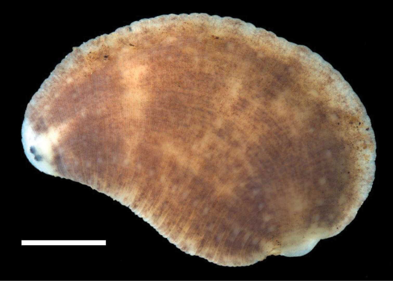
Figure 2.Dorsal surface of Placobdella appalachiensis sp. n., Holotype USNM 1232924 collected from an adult eastern hellbender (Cryptobranchus alleganiensis) from stream reach A3 in southwest Virginia, USA. Scale bar equals 1 mm.
-
Darío J. Díaz Cosín, Marta Novo, Rosa Fernández, Daniel Fernández Marchán, Mónica Gutiérrez
Zookeys
Figure 2.External view of the anterior part of the body of Eiseniona gerardoi.
-
Daniel Fernández Marchán, Rosa Fernández, Marta Novo, Darío J. Díaz Cosín
Zookeys
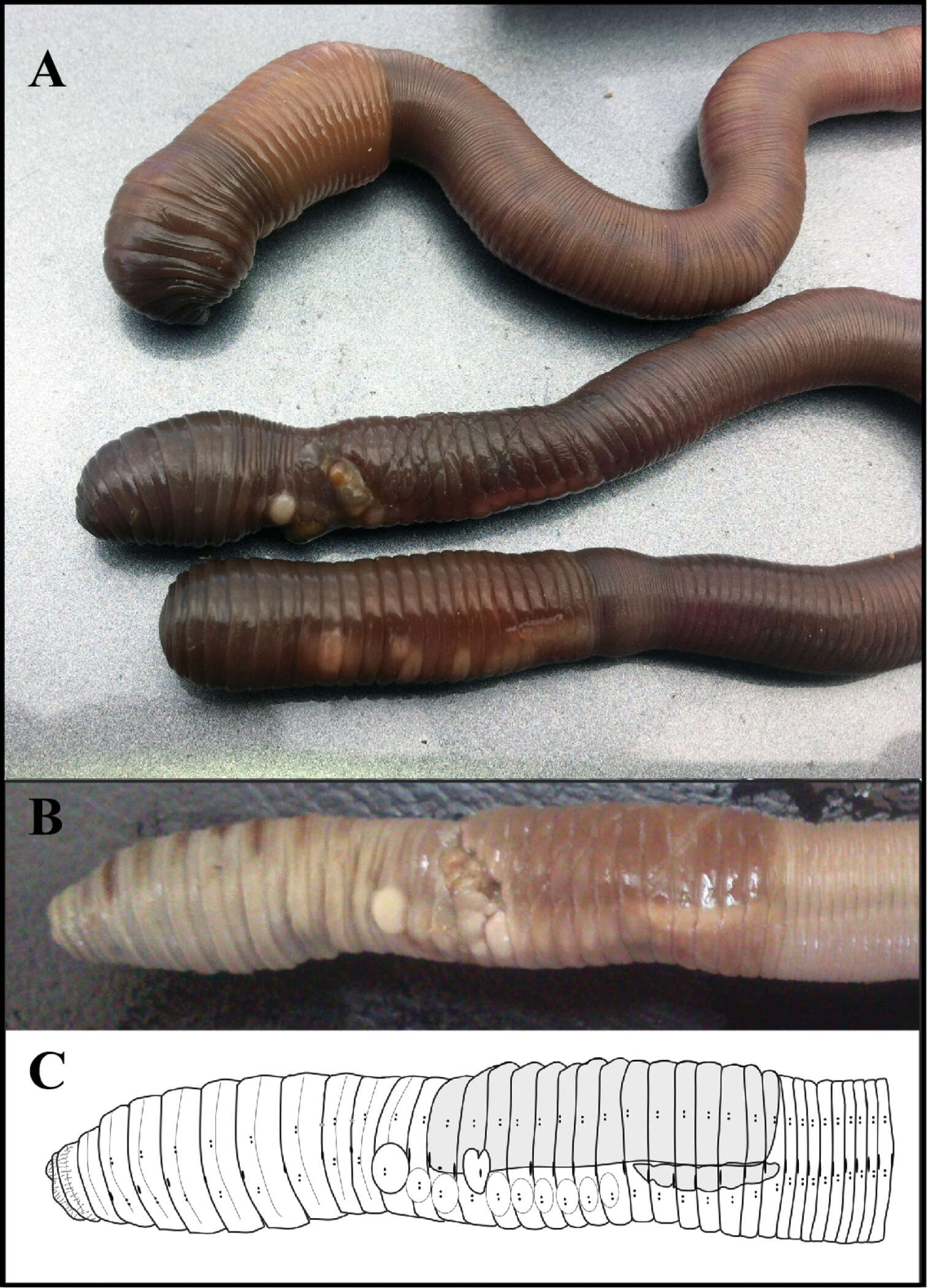
Figure 2.(A) Live specimens of Hormogaster joseantonioi sp.n. External morphology of a fixed specimen, shown in a picture (B) and diagram (C).
-
Shinri Tomioka, Eijiroh Nishi, Hiroshi Kajihara
Zookeys
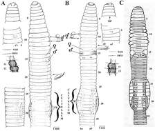
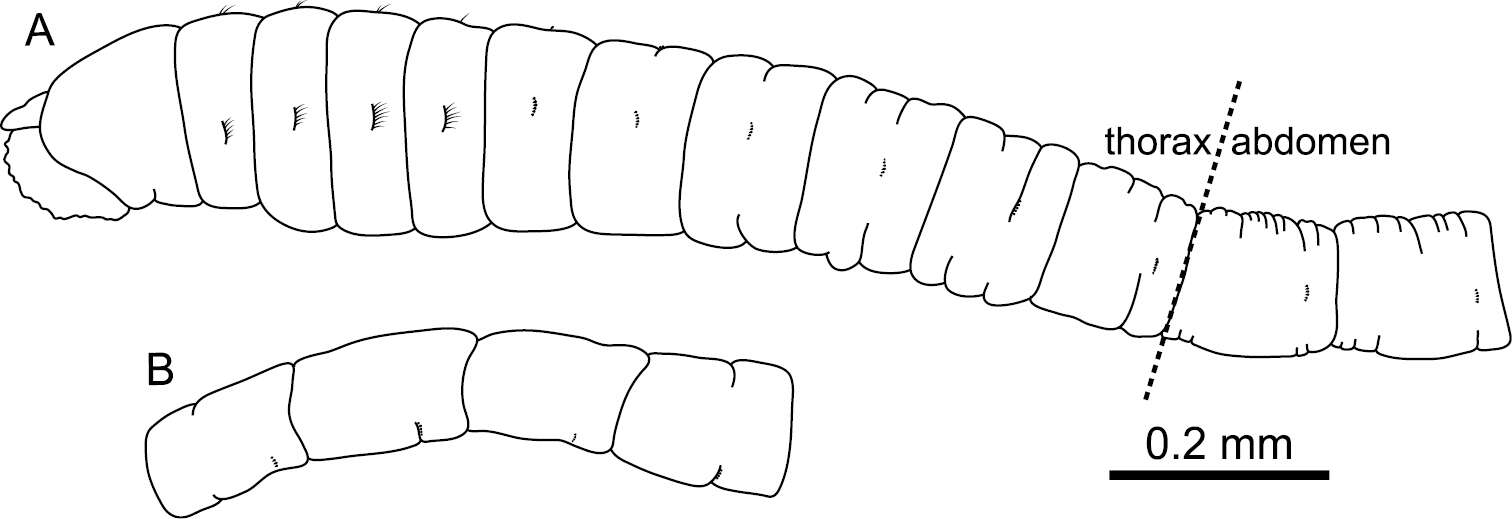
Figure 2.Mediomastus duobalteus sp. n., holotype, CBM-ZW 1088. A Anterior end of body, left lateral view B abdominal segments, left lateral view.
-
Ueangfa Bantaowong, Ratmanee Chanabun, Piyoros Tongkerd, Chirasak Sutcharit, Samuel W. James, Somsak Panha
Zookeys
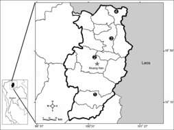
Figure 1.Map of type locality of 1 Amynthas srinan sp. n. from Srinan National Park, Nan province, 2 Amynthas phatubensis sp. n. from Tham Pha Tub Arboretum, Nan province, 3 Amynthas tontong sp. n. from Tontong Waterfall, Pua district, Nan province and 4 Amynthas borealis sp. n. from a small hill near Chaloemprakiat district, Nan province.
-
Wilsons Promontory, Victoria, Australia
-
Onna, Okinawa, Japan
-
-
Onna, Okinawa, Japan
-
Lawe Alas, Nanggroe Aceh Darussalam, Indonesia
-
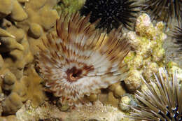
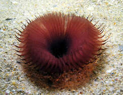
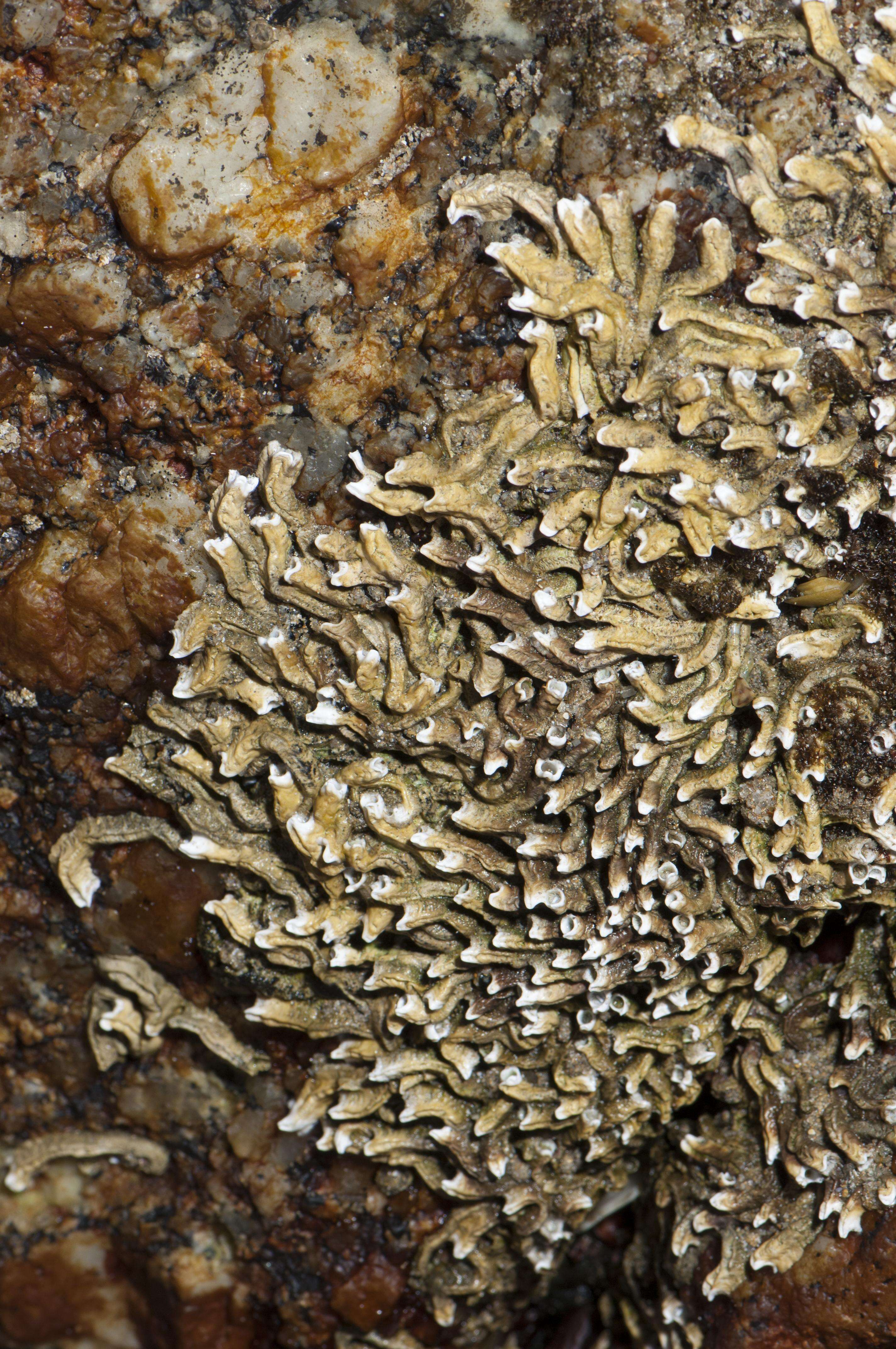
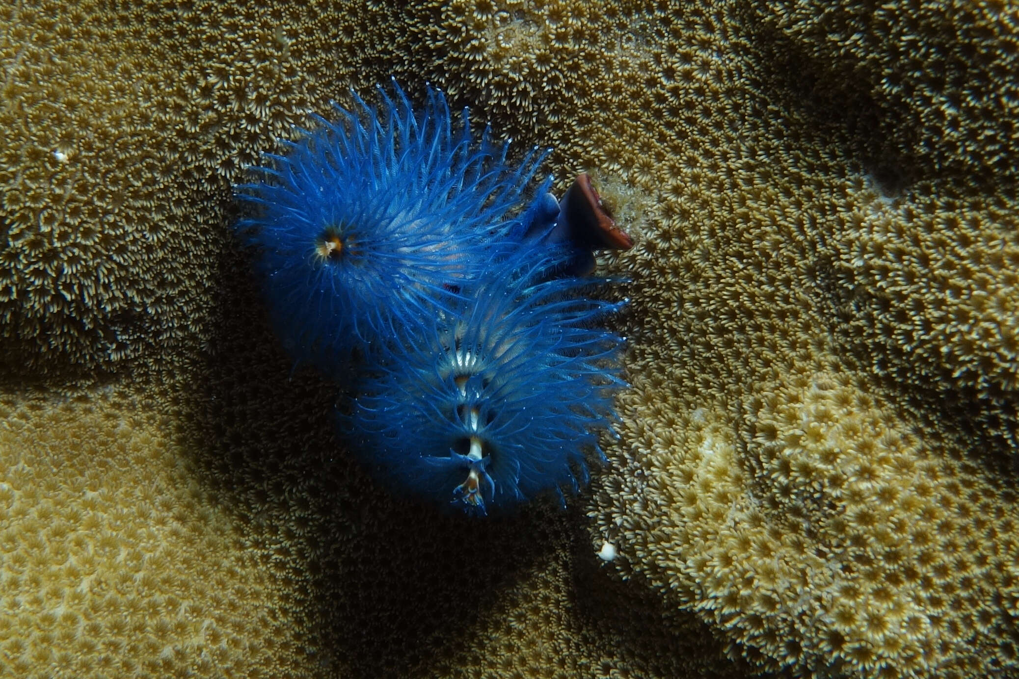
The body of the worm is buried in the sand - what you can see here is the feeding filter. They normally retract in the blink of an eye but this one was a bit slow...
-
-
-
-
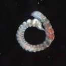
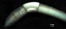
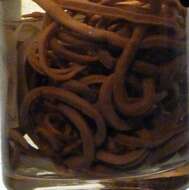
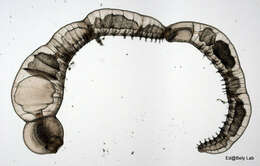
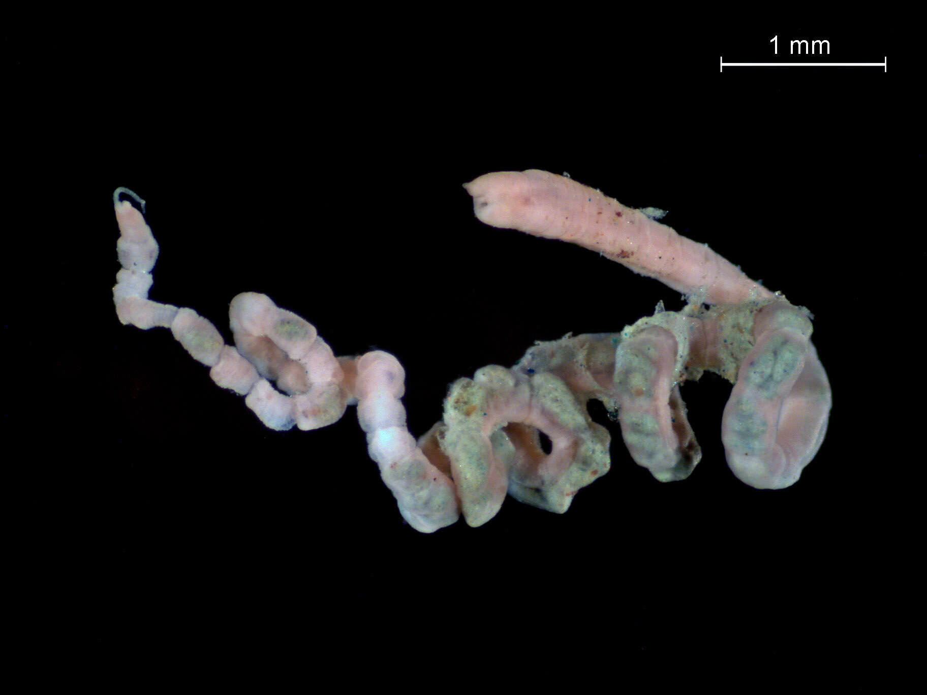
A zooid chain of Chaetogaster diaphanus, photographed live under a composite microscope.
-
-
-
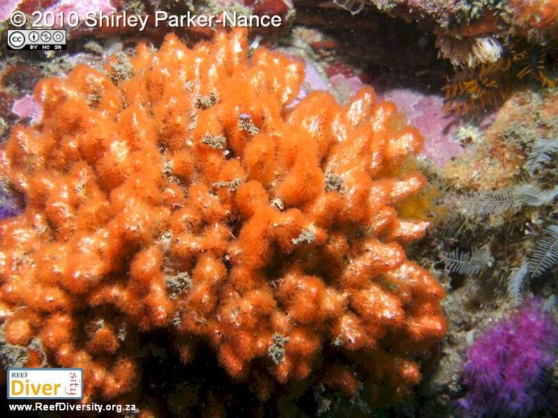
Summerstrand, Eastern Cape, South Africa