-
Chlamydomonas is a green alga, common in freshwater habitats like Swan Lake. The cells are small with two flagella used for locomotion. A single red eyespot (stigma) is used to sense light levels and control the direction of 'swimming'. Animations by Rosemary Arbur of flagellar beat patterns are available
here.
-
Scale bar indicates 50 m. Sample from the Domnental pond of Kronshagen near Kiel. The image was built up using several photomicrographic frames with manual stacking technique. Images were taken using Zeiss Axioplan with Olympus OM-D-E-M5 MKII.For permission to use of (high-resolution) images please contact postmaster@protisten.de.
-
Trieste, Friuli-Venezia Giulia, Italia
-
Rabano De Aliste, Castille and Leon, Spain
-
Caada del Hoyo, Castilla-La Mancha, Espaa
-
Cuelgamuros, Madrid, Spain
-
All Biocode files are based on field identifications to the best of the researcher’s ability at the time.
-
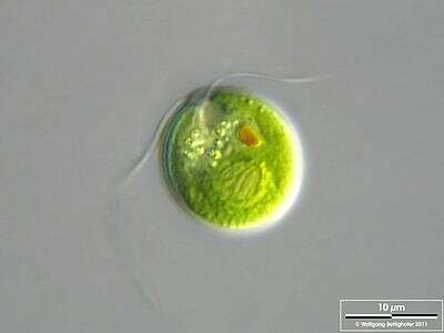
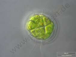
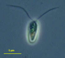
Chlamydomonas (clam-ee-doe-moan-ass), a solitary volvocid (flagellated green algal cell). Cell surrounded by a cellulosic wall, with two similar flagella emerging from near the apex. The photosynthetic pigments are located within a cup-shaped chloroplast which has a large pyrenoid with associated polysaccharide materials located posteriorly. The nucleus is located within the cup. This image shows one anterior contractile vacuole. Animations by Rosemary Arbur of flagellar beat patterns are available
here.Phase contrast.
-
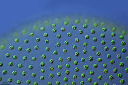
Differential interference contrast image of a cluster of cells attached to the coversl;ip by their flagella.
-
This is the flagellated form in phase contrast.
-
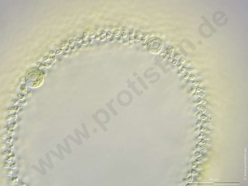
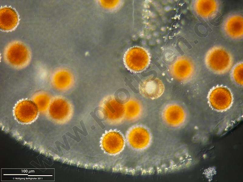
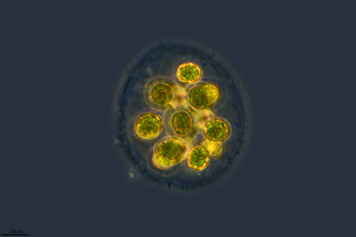
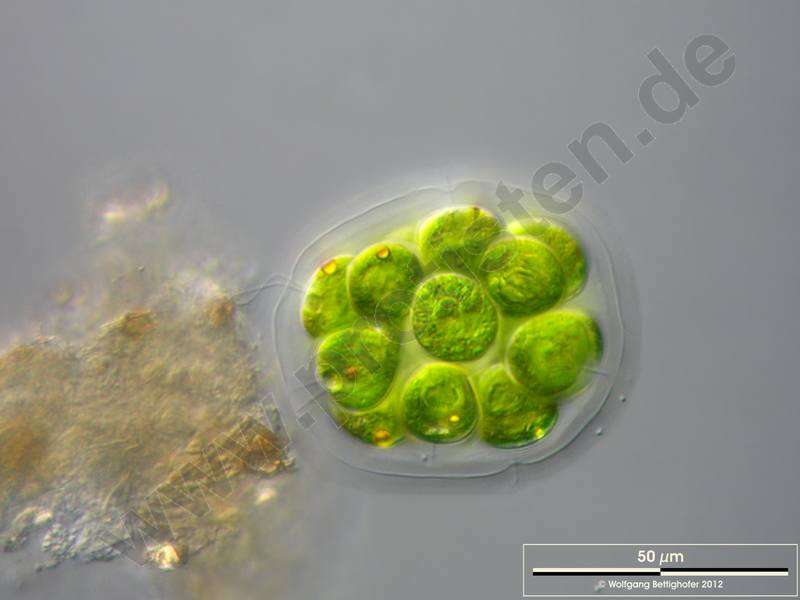
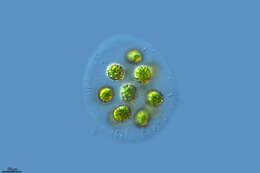
Disintegrated parent colony with zygotes. Scale bar indicates 100 m. Sample from ponds situated in the vicinity of Lake Constance. The image was built up using several photomicrographic frames with manual stacking technique. Images were taken using Zeiss Universal with Olympus C7070 CCD camera.For permission to use of (high-resolution) images please contact postmaster@protisten.de.
-
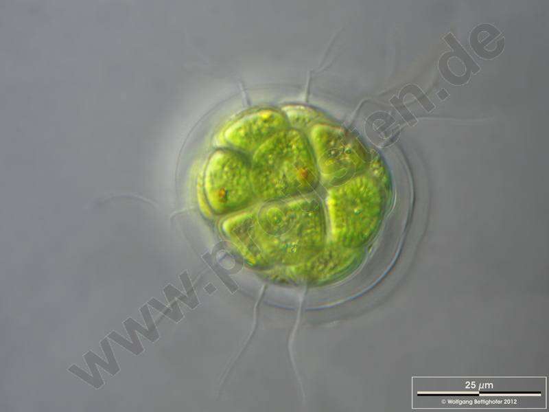
Scale bar indicates 25 m.Sample from the pond Hegne Moor situated in the vicinity of Lake Constance. The image was built up using several photomicrographic frames with manual stacking technique. Images were taken using Zeiss Universal with Olympus C7070 CCD camera.For permission to use of (high-resolution) images please contact postmaster@protisten.de.
-
Cuelgamuros, Madrid, Spain
-
All Biocode files are based on field identifications to the best of the researcher’s ability at the time.
-
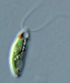
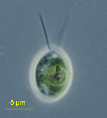
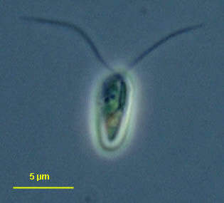
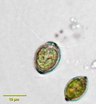
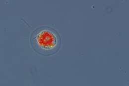
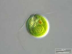
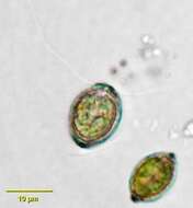
Chlamydomonas (clam-ee-doe-moan-ass), a solitary volvocid (flagellated green algal cell). Cell surrounded by a cellulosic wall. Cell damaged, no flagella. The photosynthetic pigments are located within a cup-shaped chloroplast which has a large pyrenoid with associated polysaccharide materials located posteriorly. The nucleus is located within the cup. This image shows the red eyespot to the right and two anterior contractile vacuoles. Phase contrast.
-
The scale bar indicates 10 µm. The specimen was gathered in the wetlands of Oderbruch (Oder valley 100 km north east of Berlin). The image was built up using several photomicrographic frames with manual stacking technique. Images were taken using Zeiss Universal with Olympus C7070 CCD camera.Image under Creative Commons License V 3.0 (CC BY-NC-SA).
-
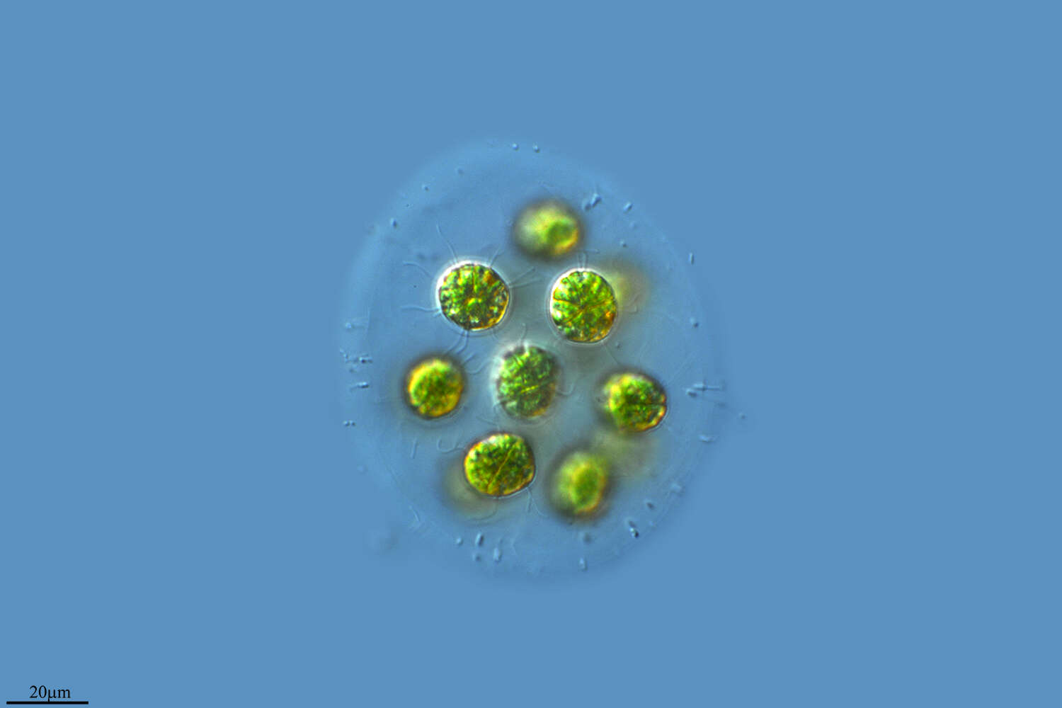
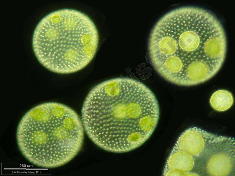
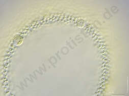
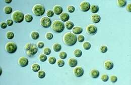
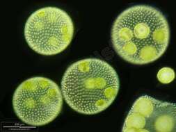
Cell colonies with daugther colonies. One parent colony disintegrates and releases daughter colonies. Scale bar indicates 250 m. Sample from a pond situated in the vicinity of Lake Constance. The image was built up using several photomicrographic frames with manual stacking technique. Images were taken using Zeiss Universal with Olympus C7070 CCD camera.For permission to use of (high-resolution) images please contact postmaster@protisten.de.
-
The specimen was gathered in the wetlands of Nationalpark Unteres Odertal ( 100 km north east of Berlin). The image was built up using several photomicrographic frames with manual stacking technique. Images were taken using Zeiss Universal with Olympus C7070 CCD camera.For permission to use of (high-resolution) images please contact postmaster@protisten.de.
-
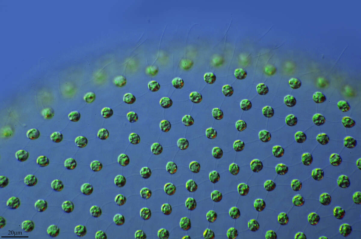
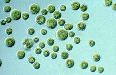
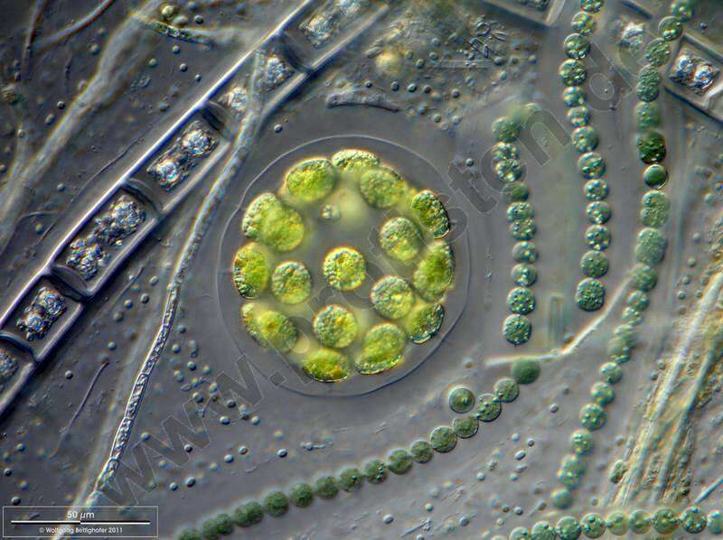
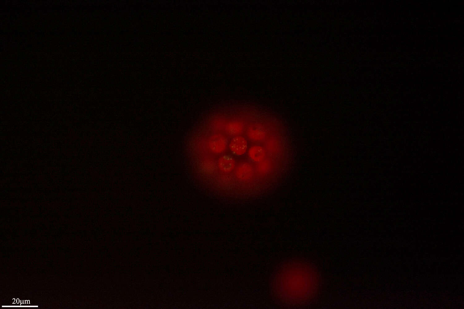
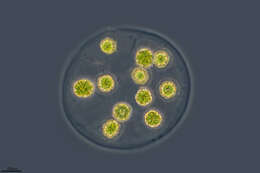
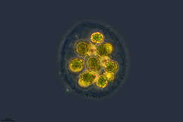
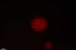
Eudorina colony accompanied by cyanobacteria (Anabaeba) and phototrophic eubacteria Pelodictyon (small bluegreen spherules embedded in mucilage). Scale bar indicates 50 m.Sample from the pond Hegne Moor situated in the vicinity of Lake Constance. The image was built up using several photomicrographic frames with manual stacking technique. Images were taken using Zeiss Universal with Olympus C7070 CCD camera.For permission to use of (high-resolution) images please contact postmaster@protisten.de.
-
All Biocode files are based on field identifications to the best of the researcher’s ability at the time.
-
Chlamydomonas (clam-ee-doe-moan-ass), a solitary volvocid (flagellated green algal cell). Cell surrounded by a cellulosic wall, with two similar flagella emerging from near the apex. Elongate species. Animations by Rosemary Arbur of flagellar beat patterns are available
here. Phase contrast.
-
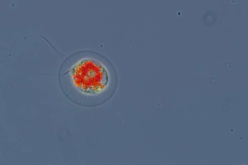
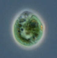
Portrait of Phacotus lenticularis, a volvocid flagellate. Lorica is composed of two shallow cup-shaped halves cemented together around the protoplast. Lorica surface is slightly rough and colored by minerals Two equal-length flagella protrude through an anterior pore in the lorica. Cup-shaped chloroplast. A stigma is present but not seen in these images. From a freshwater pond near Boise, Idaho. Brightfield.
-
Ribadelago de Franco, Castille and Leon, Spain
-
All Biocode files are based on field identifications to the best of the researcher’s ability at the time.