-
-
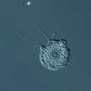
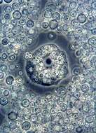
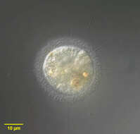
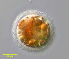
Nuclearia , nucleariid filose amoebae with thin pseduopodia. This is one of the smaller species, either N. moebiusi or N. simplex. There is a large nucleus with a dark spherical nucleolus and to the right is a contractile vacuole. The fine pseudopodia extend from the front of the cell (upper left) and are withdrawn from the back of the cell. From Lake Donghu, China. Phase contrast micrograph.
-
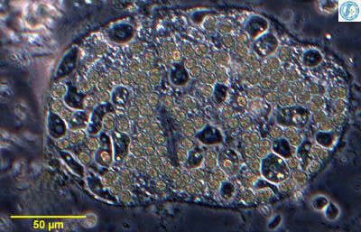
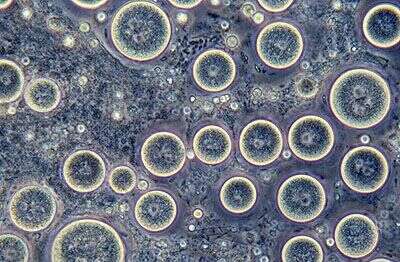
Nuclearia , nucleariid filose amoebae with thin pseduopodia. This is one of the smaller species, either N. moebiusi or N. simplex. It has invaded a colony of Microcystis (cyanobacteria) and is eating the bacterial cells and multiplying within the colony. From Lake Donghu, China Phase contrast micrograph.
-
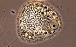
Nuclearia, one of the naked filose amoebae, producing thin pseudopodia without internal skeletons. This is one of the larger more rounded species, eats detritus and algae. Phase contrast micrograph.
-
Nuclearia isolated from sandy sediments from Little Sippiwissett salt marsh. Micrograph taken by Jeffrey Cole.
-
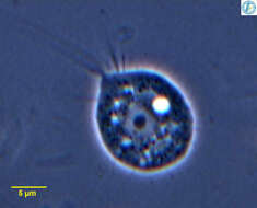
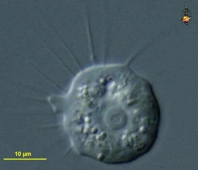
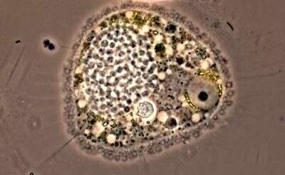
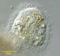
Nuclearia (new-clee-air-ee-a), a nucleariid (cristi-discoid) filose amoeba. Exclusively with thin pseudopodia without skeletal supports. More of these project from the front of the cell (to the left), whereas they appear a little more crumpled at the posterior end. The structure slightly right and below the centre is the nucleus, with a large nucleolus and the nucleolus having a small empty central region. Eats bacteria, algae, detritus. Differential interference contrast.
-
This cell, like many Nuclearia species, has been consuming algae. The nucleus is the clear area with the dark nucleolus in the center. Nucleariid amoebae have thin non-stiff pseudopodia. Phase contrast image.
-
Cysts, from a culture. Phase contrast.
-
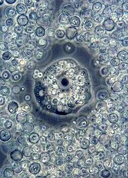
Live cell, phase contrast. The fine pseudopodia are typical of nuclearid filose amoebae. The surrounding material is mostly comprised of yeast cell walls (the species is provided with yeast as food).
-
-
-
-
-
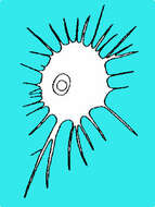
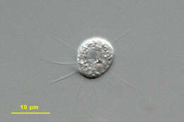
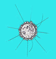
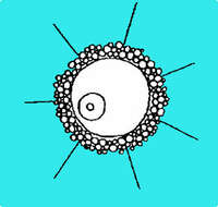
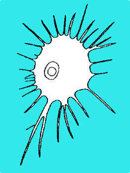
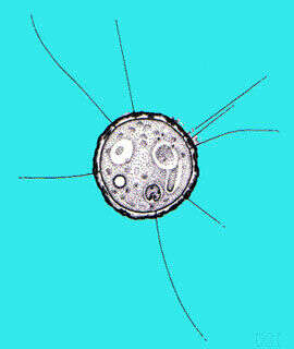
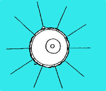
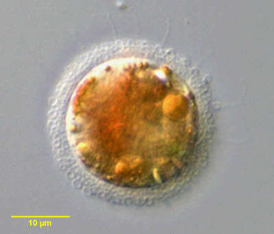
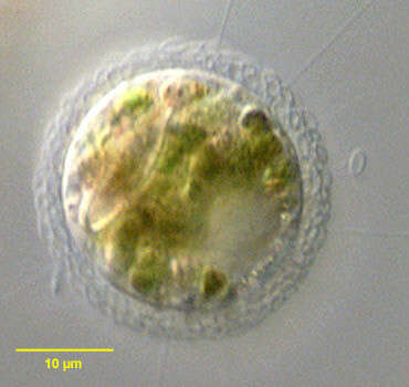
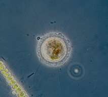
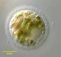
Pompholyxophrys is a filose amoeba. Its cell surface is covered by a layer of delicate siliceous (glass-like) spheres. It has thin, delicate pseudopodia that it uses for movement. The structure to the right in the cell is its nucleus.
-
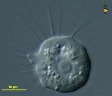
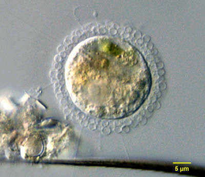
Portrait of Pompholyxophrys, one of the heliozoon-like amoebae previously assigned to the order Rotosphaerida. Pompholyxophrys has a periplast composed of endogenously formed siliceous elements of a single type within a species. These are spherical in the type species P. punicea but may be ovoid (see images of P. ovuligera), discoid or bone-shaped. Radiating spicules are absent. What appear at first glance to be axopodia are, in fact, filopods that lack axonemes and extrusomes. Contracted filopodia may appear granular leading to confusion but close examination of the extended filopodia shows extrusomes are absent. From standing freshwater Typha latifolia marsh near Boise, Idaho. Differential interference contrast
-
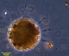
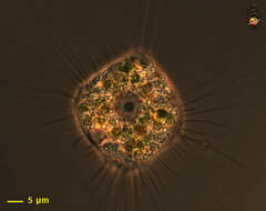
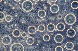
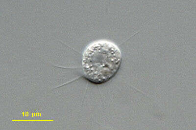
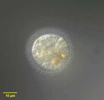
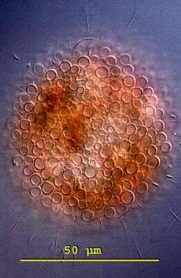
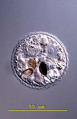
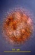
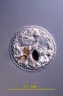
Pompholyxophrys (pom-folly-zoff-riss) punicea. The cytoplasm of the spherical body is colorless or reddish, often interspersed with colored granules and green or brown food particles. The outer periplast is built up from conspicuous minute colourless spherical granules (perles) arranged in concentric layers. The granules are glass like hollow spheres and arranged in concentric layers to form a compact envelope. The large nucleus is located eccentricly. The straight and pointed pseudopodia are tenuous and indistinct. Individuals are found occasionally in ponds and swamps. This specimen was collected in a bog pond near Konstanz, Germany. The outer sphere of colourless spherical perles can be seen to be lying in concentric layers. Differential interference contrast.
-
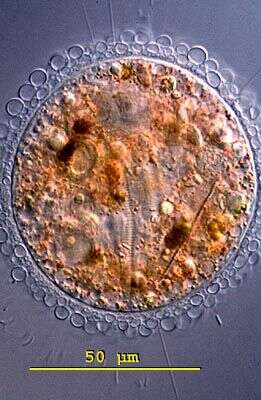
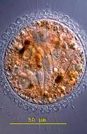
Pompholyxophrys (pom-folly-zoff-riss) punicea. The cytoplasm of the spherical body is colorless or reddish, often interspersed with colored granules and green or brown food particles. The outer periplast is built up from conspicuous minute colourless spherical granules (perles) arranged in concentric layers. The granules are glass like hollow spheres and arranged in concentric layers to form a compact envelope. The large nucleus is located eccentricly. The straight and pointed pseudopodia are tenuous and indistinct. Individuals are found occasionally in ponds and swamps. This specimen was collected in a bog pond near Konstanz, Germany. The focal plane on the surface of the sphere of spherical granules. Each perle measures 3-4 microns in diameter. Differential interference contrast.
-
Portrait of Pompholyxophrys, one of the heliozoon-like amoebae previously assigned to the Rotosphaerida. Pompholyxophrys has a periplast composed of endogenously formed siliceous elements of a single type within a species. These are spherical in the type species P. punicea but may be ovoid (see images of P. ovuligera), discoid or bone-shaped. Radiating spicules are absent. What appear at first glance to be axopodia are, in fact, filopods which lack axonemes and extrusomes. Contracted filopodia may appear granular leading to confusion but close examination of the extended filopodia shows extrusomes are absent. From standing freshwater near Boise, Idaho. Differential interference contrast.
-
Detail of Pompholyxophrys, one of the heliozoon-like amoebae previously assigned to the order Rotosphaerida. Pompholyxophrys has a periplast composed of endogenously formed siliceous elements of a single type within a species. These are spherical in the type species P. punicea but may be ovoid (see images of P. ovuligera), discoid or bone-shaped. Radiating spicules are absent. This individual has completely retracted its filopodia. From standing freshwater near Boise, Idaho. Differential interference contrast
-
Portrait of Pompholyxophrys, one of the heliozoon-like amoebae previously assigned to the order Rotosphaerida. Pompholyxophrys has a periplast composed of endogenously formed siliceous elements of a single type within a species. These are spherical in the type species P. punicea but may be ovoid (see images of P. ovuligera), discoid or bone-shaped. Radiating spicules are absent. What appear at first glance to be axopodia are, in fact, filopods that lack axonemes and extrusomes. Contracted filopodia may appear granular leading to confusion but close examination of the extended filopodia shows extrusomes are absent. This image shows the eccentric nucleus at approximately 12 o clock in the upper half of the cell. From standing freshwater Typha latifolia marsh near Boise, Idaho. Differential interference contrast.
-
Pompholyxophrys punicea is a filose amoeba, the cell body of which is surrounded by silceous perles, hollow beads with performated siliceous walls. The amoebae tend to eat detritus and algae. Has been subject of a talk-radio quiz - spell Pompholyxophrys punicea and win the glorious prize of an ICOP-X t-shirt. No-one won.
-
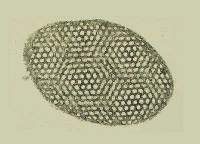
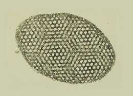
Transmission electron micrograph of a whole mount of a siliceous perle from the surface of the filose amoeba.
-
Pinaciophora (pin-ass-ee-off-or-a). The cytoplasm of the spherical body is colorless or reddish, often interspersed with colored granules and green or brown food particles. The outer periplast is built up from minute colourless spherical plates. The large nucleus is located eccentricly. The straight and pointed pseudopodia are tenuous and indistinct. Individuals are found occasionally in ponds and swamps. This specimen was collected in a bog pond near Konstanz, Germany. Differential interference contrast. T
-
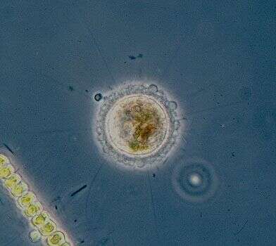
Portrait of Pompholyxophrys ovuligera, one of the heliozoon-like amoebae previously assigned to the order Rotosphaerida. Pompholyxophrys has a periplast composed of endogenously formed siliceous elements of a single type within a species. These are spherical in the type species P. punicea but may be ovoid (as seen here), discoid or bone-shaped. Radiating spicules are absent. A detached (American) football-shaped scale is seen on your right in this image. What appear at first glance to be axopodia are, in fact, filopods protruding between the periplast scales. The filopods lack axonemes and extrusomes. Contracted filopodia may appear granular leading to confusion but close examination of the extended filopodia shows extrusomes are absent. This individual has been feeding on algae and diatoms. From standing freshwater near Boise, Idaho