-
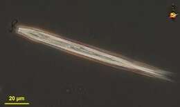
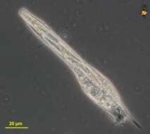
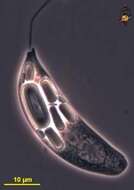
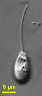
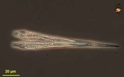
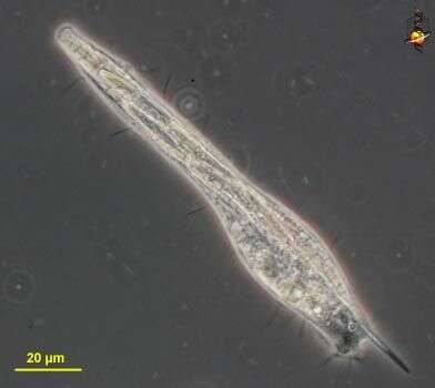
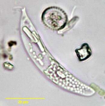
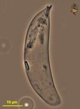
Cyclidiopsis (sigh-clid-ee-op-sis) is a colourless heterotrophic euglenid, but is unusual in that it has an eyespot. Elongate, anterior slightly trumpet-like with one emergent flagellum. Not often encountered. This is a dividing cell. Phase contrast.
-
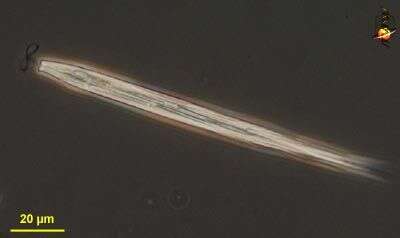
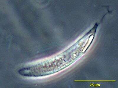
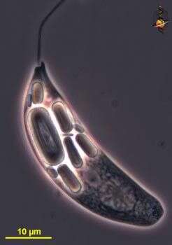
Cyclidiopsis (sigh-clid-ee-op-sis) is a colourless heterotrophic euglenid, but is unusual in that it has an eyespot. Elongate, anterior slightly trumpet-like with one emergent flagellum. This image is to show the beat pattern of the flagellum. Not often encountered. Phase contrast.
-
Cyclidiopsis (sigh-clid-ee-op-sis) is a colourless heterotrophic euglenid, but is unusual in that it has an eyespot. Elongate, anterior slightly trumpet-like with one emergent flagellum. Not often encountered. This cell has just been eaten by a ciliate. Phase contrast.
-
-
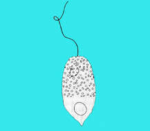
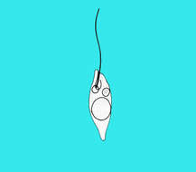
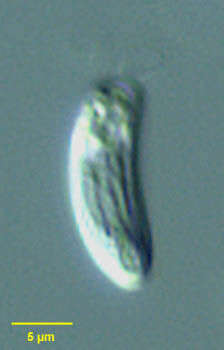
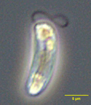
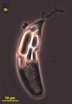
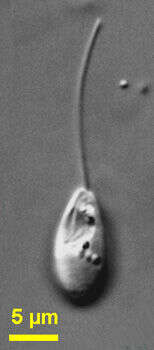
Rhabdomonas, a swimming colourless euglenid. One flagellum emerges from near the anterior apex of the cell and beats in a figure-of-eight pattern. Surface with slightly spiralling ridges. Differential interference contrast optics.
-
Rhabdomonas, a swimming colourless euglenid. One flagellum emerges from near the anterior apex of the cell and beats in a figure-of-eight pattern. Surface with slightly spiralling ridges. Phase contrast.
-
Rhabdomonas spiralis (PRINGSHEIM,1942).Collected from a freshwater pond near Boise, Idaho.July 2007. DIC.
-
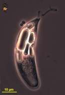
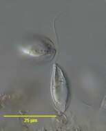
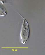
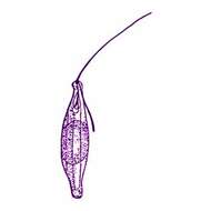
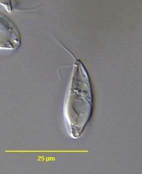
Portrait of Menoidium a colorless euglenoid flagellate. Strongly flattened. One side curved with the other more straight. One emergent flagellum. Stigma absent. Paramylon bodies are dimorphic with smaller round and larger elongate ring forms. Swims rotating on long axis. Highly refractile. From standing freshwater near Boise, Idaho. Brightfield.
-
Portrait of Menoidium a colorless euglenoid flagellate. Strongly flattened. One side curved with the other more straight. One emergent flagellum. Stigma absent. Paramylon bodies are dimorphic with smaller round and larger elongate ring forms. Swims rotating on long axis. From standing freshwater near Boise, Idaho. Phase contrast.
-
-
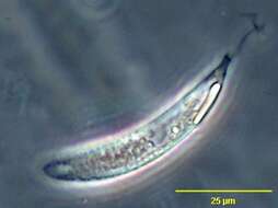
Menoidium pellucidum. Cell observed in freshwater habitats in the vicinity of Broome, Western Australia in September 2003. This image was taken using phase contrast optics. This work was supported by the Australian Biological Resources Study.
-
Menoidium pellucidum. Empty pellicle observed in freshwater habitats in the vicinity of Broome, Western Australia in September 2003. This work was supported by the Australian Biological Resources Study.
-
Menoidium pellucidum. Cell observed in freshwater habitats in the vicinity of Broome, Western Australia in September 2003. This image was taken using phase contrast optics. This work was supported by the Australian Biological Resources Study.
-
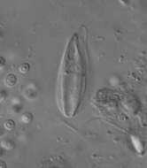
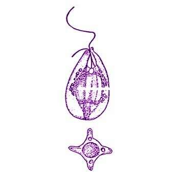
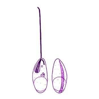
Sphenomonas quadrangularis Stein, 1878. Biflagellated, gliding euglenid, rigid, 17 - 30 microns (average 23.0 microns) long and 8 - 15 microns (average 11.5 microns) wide. Cell shape ellipsoid, anterior end of cell pointed, apically slightly oblique, posterior end of cell broadly rounded. Pellicle hyaline with 4 longitudinal strongly developed keels, each containing a series of small paramylon grains. In cruciate the cell shape looks square with concave sides and roundish angles. The anterior flagellum is about one or one and a half of the cell length and the recurrent flagellum about one quarter to one thirth of the cell length. The centre of cell always contains a large hyaline inclusion. The reservoir is situated in mid-line in the first quarter of the cell with an associated contractile vacuole.
-
Portrait of the colorless euglenid flagellate, Sphenomonas quadrangularis (Playfair, 1921).DIC.
-
Portrait of the colorless euglenid flagellate, Sphenomonas quadrangularis (Playfair, 1921).DIC.
-
Portrait of the colorless euglenid flagellate, Sphenomonas quadrangularis (Playfair, 1921).DIC.
-
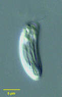
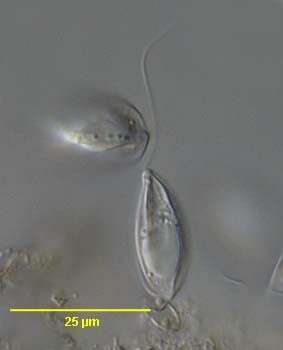
Sphenomonas (sfeen-owe-moan-ass) angusta Skuja, 1956. Cells are 10 to 14 microns long, 4 to 7 microns wide, not flattened and with a dorsal groove. The cells are anteriorly obliquely truncated and posteriorly rounded. The cell bodies are slightly curved: the right margin of the cell is straighter than the left one. With two flagella, unequal in length, emerging from a relatively large flagellar pocket located in the right anterior end of the cell. The anterior flagellum is about 1.5 times cell length and its proximal part moves actively, the trailing posterior flagellum is less than 0.5 times the cell length. One large refractile inclusion often occupies the posterior part of the cell. Commonly observed.
-
Sphenomonas angusta Skuja, 1956. Cells are 10 to 14 microns long, 4 to 7 microns wide, not flattened and with a dorsal groove. The cells are anteriorly obliquely truncated and posteriorly rounded. The cell bodies are slightly curved: the right margin of the cell is straighter than the left one. With two flagella, unequal in length, emerging from a relatively large flagellar pocket located in the right anterior end of the cell. The anterior flagellum is about 1.5 times cell length and its proximal part moves actively, the trailing posterior flagellum is less than 0.5 times the cell length. One large refractile inclusion often occupies the posterior part of the cell.
-
-
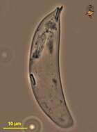
Sphenomonas teres (Stein, 1878) Klebs, 1893. Rigid, gliding euglenid, cell shape elongate elliptical, anterior end of cell narrow, apically oblique with slight neck and reflexed collar. Cell is about 11 - 30 microns long. Posterior end of cell also narrow, slightly pointed.or papillated. Two flagella insert in the flagellar pocket, unequal in length. The anterior flagellum is about the same length as the cell or slightly longer. The second flagellum is about one quarter the length of the cell. The flagellar pocket is located dorsally and has an associated contractile vacuole. Pellicle with at least 6-8 delicate, longitudinal ridges, spaced about 1.5 - 2 microns Typically there is a large hyaline inclusion of cell located more or less in the posterior part of cell. Cell contain a few small roundish paramylon grains in the posterior part of cell and some more in the anterior part of cell. T Clostenema sociale Stokes synomised by Huber-Pestalozzi, 1955
-
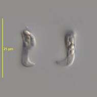
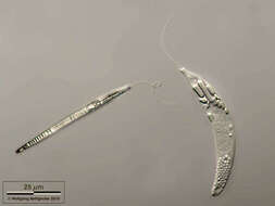
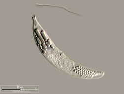
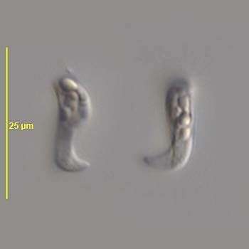
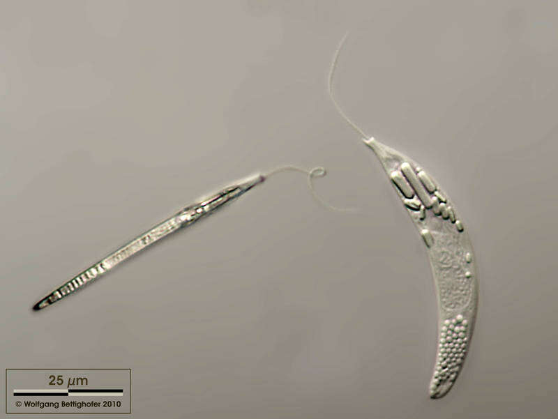
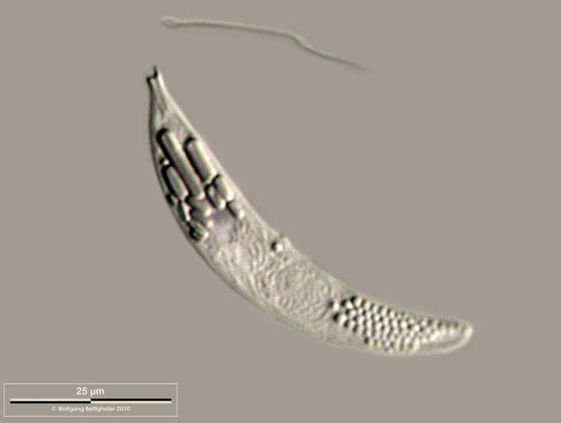
Sampling date 09/2009. Scale bars indicate 25 µm.Two images.First:Top and lateral view of same cell.Second:There are several paramylon bodies in the apical region, the nucleus is in the center of the cell.Please click on < or > on the image edges or on the dots at the bottom edge of the images to browse through the slides!Place name: Bog Hegne Moor near Lake Constance (Germany) Latitude: 47.718106 Longitude: 9.093974Microscope Zeiss Universal, camera Olympus C7070WZ. DOF images.© Wolfgang Bettighofer,images under Creative Commons License V 3.0 (CC BY-NC-SA).For permission to use of (high resolution) images please contact
postmaster@protisten.de.For further information about the image, please click here:
Link to protisten.de page
-
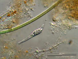
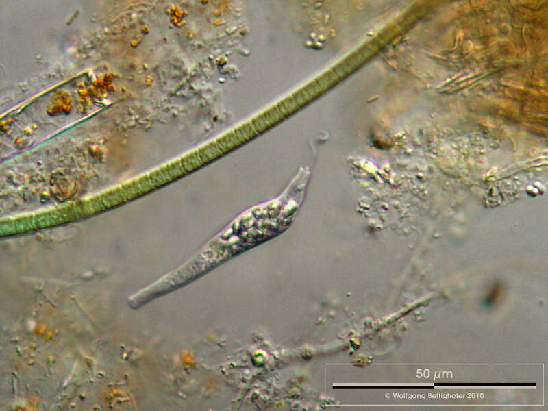
Sampling date 09/2009. Scale bars indicate 50 µm.Two images.First:Euglenid without chloroplasts, highly metabolic, two flagella, one of them (short one) sometimes trailing.Second:Motion study.Please click on < or > on the image edges or on the dots at the bottom edge of the images to browse through the slides!Place name: Bog Hegne Moor near Lake Constance (Germany) Latitude: 47.718106 Longitude: 9.093974Microscope Zeiss Universal, camera Olympus C7070WZ. DOF images.© Wolfgang Bettighofer,images under Creative Commons License V 3.0 (CC BY-NC-SA).For permission to use of (high resolution) images please contact
postmaster@protisten.de.For further information about the image, please click here:
Link to protisten.de page