-
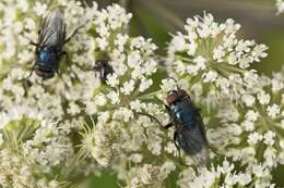
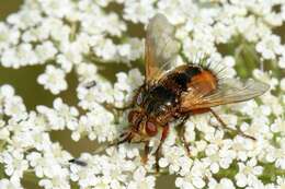
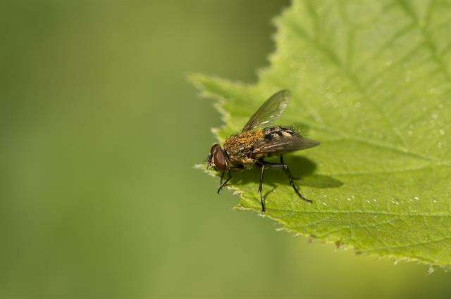
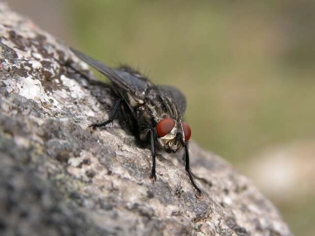
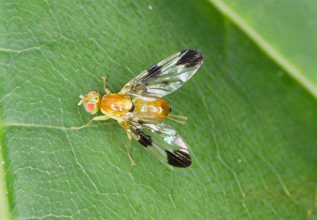
Kværkeby Mose, Midtsjælland, Denmark
-
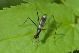
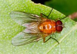
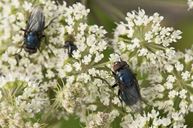
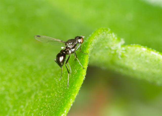
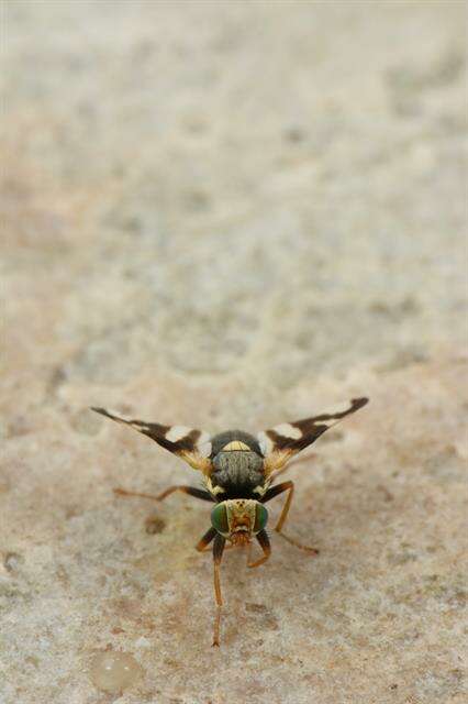
Kværkeby Mose, Midtsjælland, Denmark
-
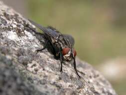
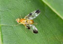
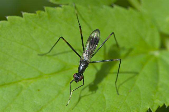
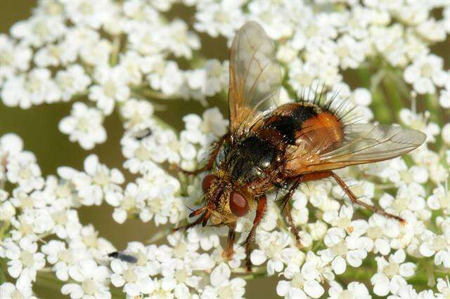
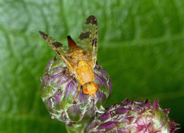
Kværkeby Mose, Midtsjælland, Denmark
-
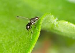
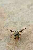
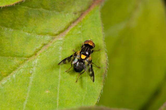
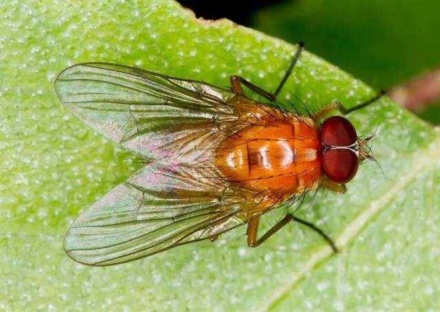
Kværkeby, Midtsjælland, Denmark
-
Allindelille Fredskov ved Ringsted, Denmark
-
Hobro
-
Asker, Akershus, Norge
-
Asker, Akershus, Norge
-
Hostrup grusgrav, Hobro, Jylland, Danmark
-
Asker, Akershus, Norge
-
Asker, Akershus, Norge
-
Jernhatten
-
Asker, Akershus, Norge
-
Centers for Disease Control/Division of Parasitic Diseases and Malaria
EOL staff
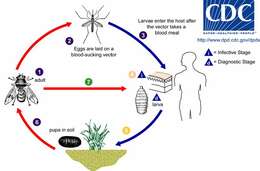
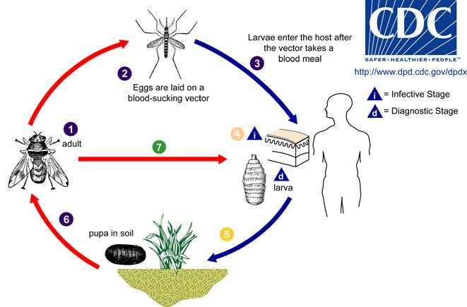
Generalized life cycle of flies causing myiasis in humans based on Human Bot Fly (click on image for description of variations)Adults of the Human Bot Fly Dermatobia hominis are free-living flies (1). Adults capture blood-sucking arthropods (such as mosquitoes) and lay eggs on their bodies, using a glue-like substance for adherence (2). Bot fly larvae develop within the eggs, but remain on the vector until it takes a blood meal from a mammalian or avian host. Newly-emerged bot fly larvae then penetrate the host's tissue (3). The larvae feed in a subdermal cavity (4) for 5-10 weeks, breathing through a hole in the host's skin. Mature larvae drop to the ground (5) and pupate in the environment. Larvae tend to leave their host during the night and early morning, probably to avoid desiccation. After approximately one month, the adults emerge (6) to mate and repeat the cycle. Other genera of myiasis-causing flies (including Cochliomyia, Cuterebra, and Wohlfahrtia) have a more direct life cycle, where the adult flies lay their eggs directly in, or in the vicinity of, wounds on the host (7). In Cochliomyia and Wohlfahrtia infestations, larvae feed in the host for about a week, and may migrate from the subdermis to other tissues in the body, often causing extreme damage in the process.From
Centers for Disease Control Parasites and Health website
-
Pest and Diseases Image Library, Bugwood.org
EOL staff
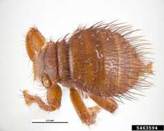
Braula coeca
Descriptor: Adult(s) Image type: Museum Image view: Dorsal / Abaxial / Back Image location: Australia Photographer Information Name:
Pest and Diseases Image Library Country: Australia - See more at: http://www.insectimages.org/browse/detail.cfm?imgnum=5463594#sthash.reR8DOwB.dpuf
-
Xiao-Yan Liu, Ding Yang, Emilia P. Nartshuk
Zookeys
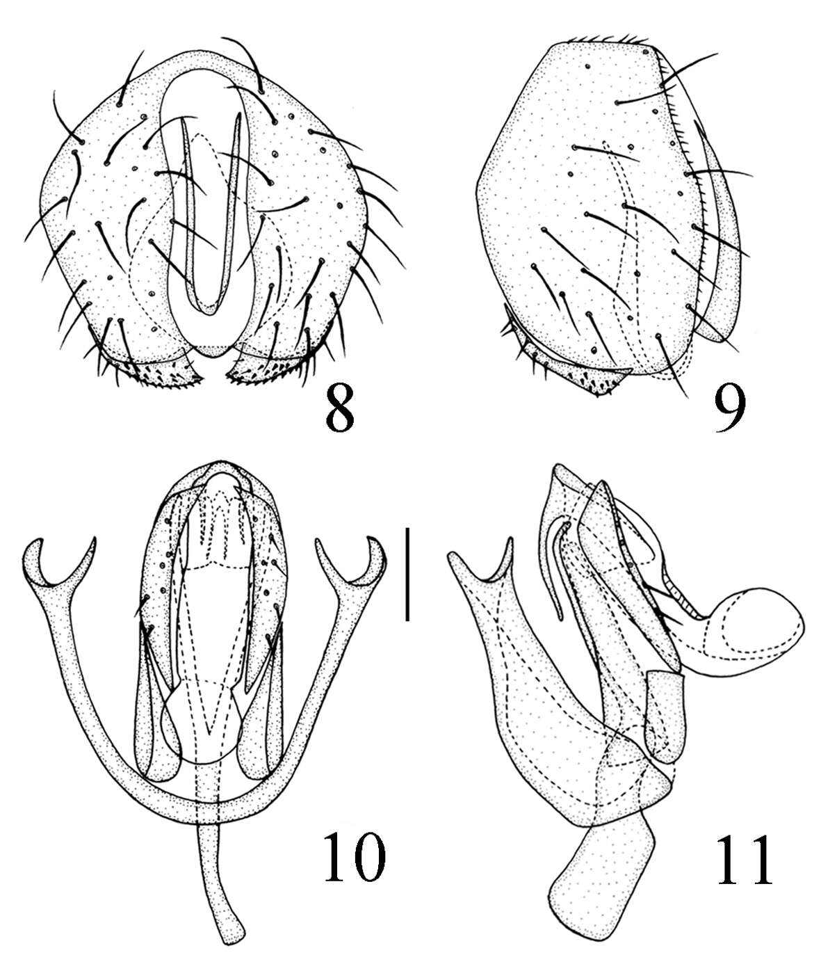
Figures 8–11. Thressa bimaculata sp. n., male. 8 epandrium, posterior view 9 epandrium, lateral view 10 hypandrium and phallic complex, ventral view 11 hypandrium and phallic complex, lateral view. Scale bar = 0.05mm.
-
Saeed Mohamadzade Namin, Jamasb Nozari
Zookeys
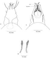
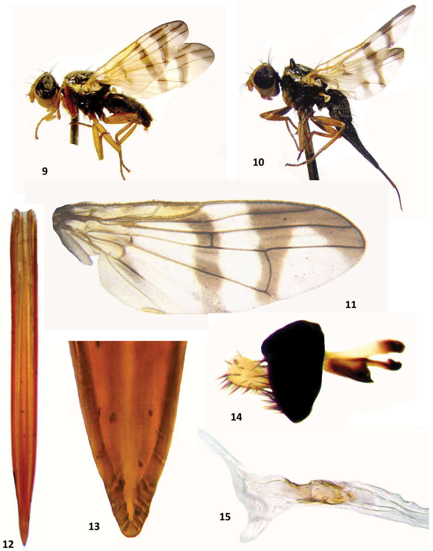
Figures 9–15. Urophora merzi sp. n., 9 ♂, total view, left 10 ♀ (Holotype), total view, left 11 wing pattern (Holotype) 12 aculeus 13 aculeus tip 14 epandrium 15 male terminalia.
-
Junhua Zhang, Ding Yang, Wayne N. Mathis
Zookeys
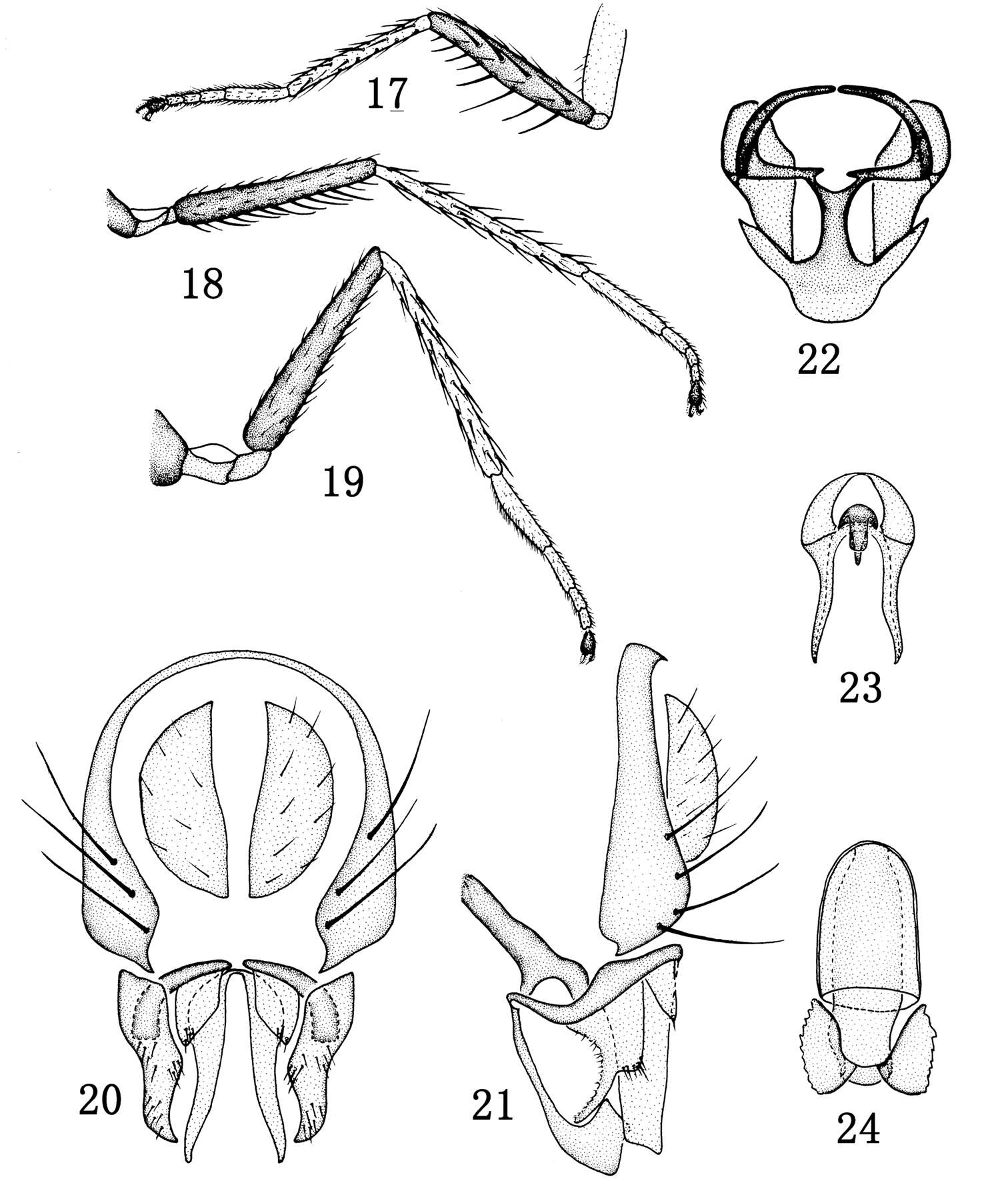
Figures 17–24.Rhynchopsilopa jinxiuensis sp. n. (male) 17 foreleg 18 midleg 19 hindleg 20 terminalia (epandrium, cercus, presurstyli, postsurstyli, aedeagus), posterior view 21 terminalia (epandrium, cercus, presurstylus, postsurstylis, aedeagus, phallapodeme, gonite/subepandrial plate, hypandrium), lateral view 22 terminalia (surstyli, gonite/subepandrial plate, hypandrium), ventral view 23 aedeagus and phallapodeme, ventral view. (female) 24 Ventral receptacle.
-
Wayne N. Mathis, Tadeusz Zatwarnicki
Zookeys
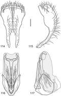
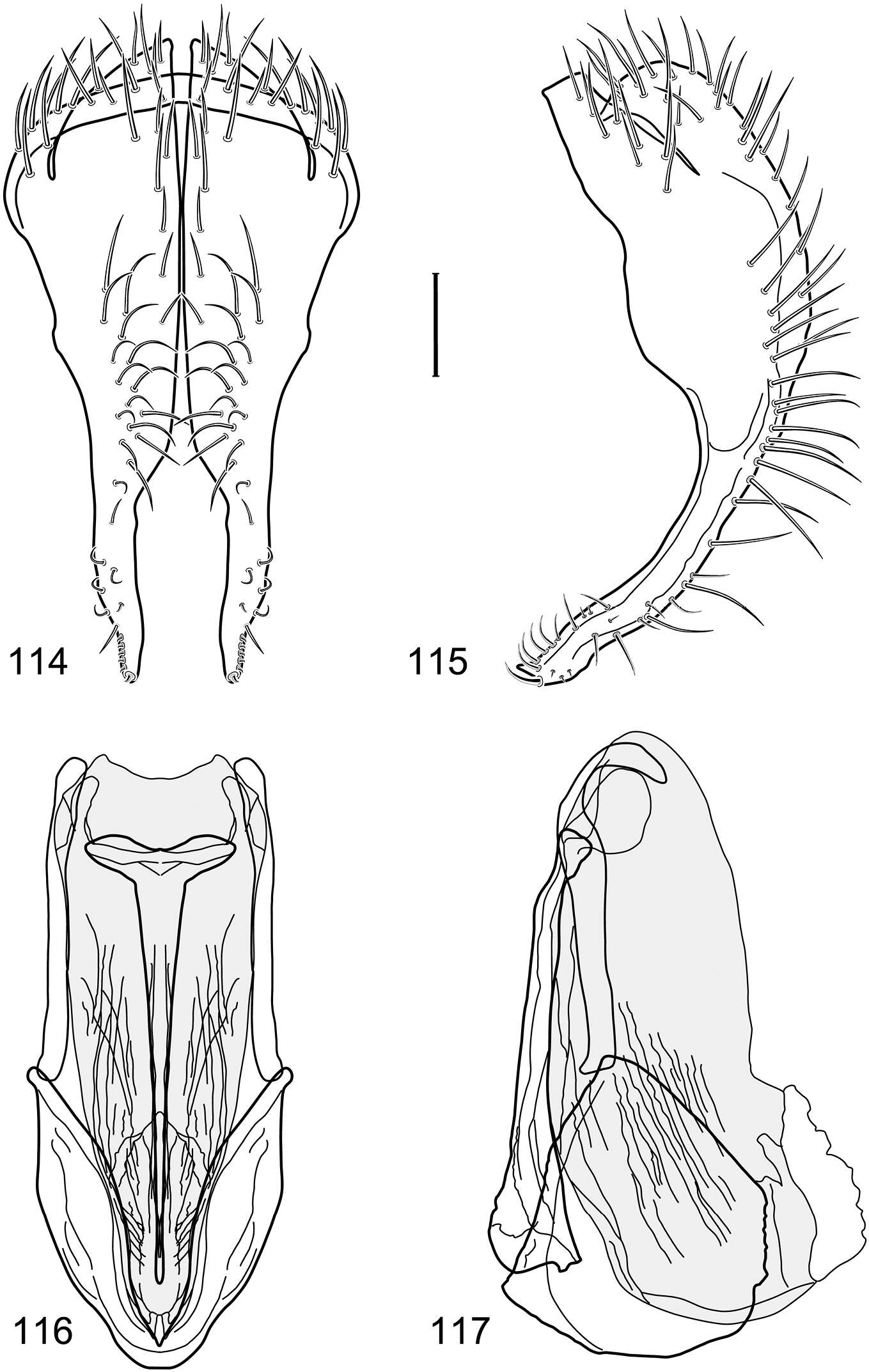
Figures 114–117.Illustration of Polytrichophora prolata sp. n. (male) (Belize. Stann Creek: Cockscomb Basin Wildlife Sanctuary) 114 epandrium and cerci, posterior view 115 same, lateral view 116 internal structures of male terminalia (aedeagus [shaded], phallapodeme, gonite, hypandrium), ventral view 117 same, lateral view. Scale bar = 0.1 mm.
-
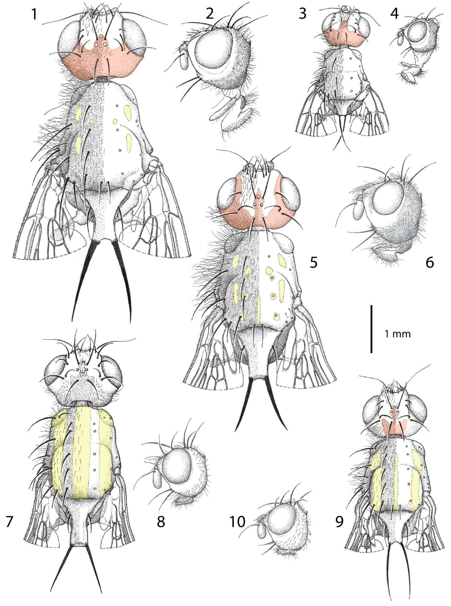
Maurizio Mei, Daniel Whitmore, Giuseppe Lo Giudice, Pierfilippo Cerretti
Zookeys
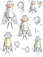
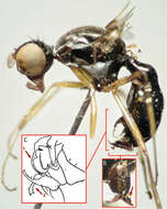
Figures 1−10.Males of Centrophlebomyia spp. 1, 3, 5, 7, 9 head and thorax in dorsal view 2, 4, 6, 8, 10 head in lateral view 1−4 Centrophlebomyia anthropophaga (Italy) 5−6 Centrophlebomyia furcata (Italy) 7−8 Centrophlebomyia grunini (Russian Far East) 9−10 Centrophlebomyia orientalis (India). In red the microtomentum pattern of head; in yellow the shiny, non microtomentose, pattern of thorax.
-
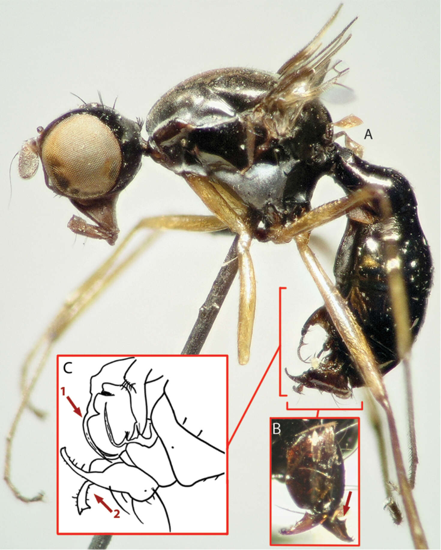
Yuchen Ang, Ling Jing Wong, Rudolf Meier
Zookeys
Figure 4.Images of holotype (A, B) and drawing (C) from description for Perochaeta orientalis, male. A Image of habitus, lateral view B Image of hypopygium, dorsal view; red arrow pointing to the median protrusion on the surstylus C Drawing of abdominal posterior (lateral view) as reproduced from Duda (1926); red arrow 1 shows how illustration has fused the two setae into one, red arrow 2 shows how the drawing fails to display the median protrusion as seen in Fig. 1G.
-
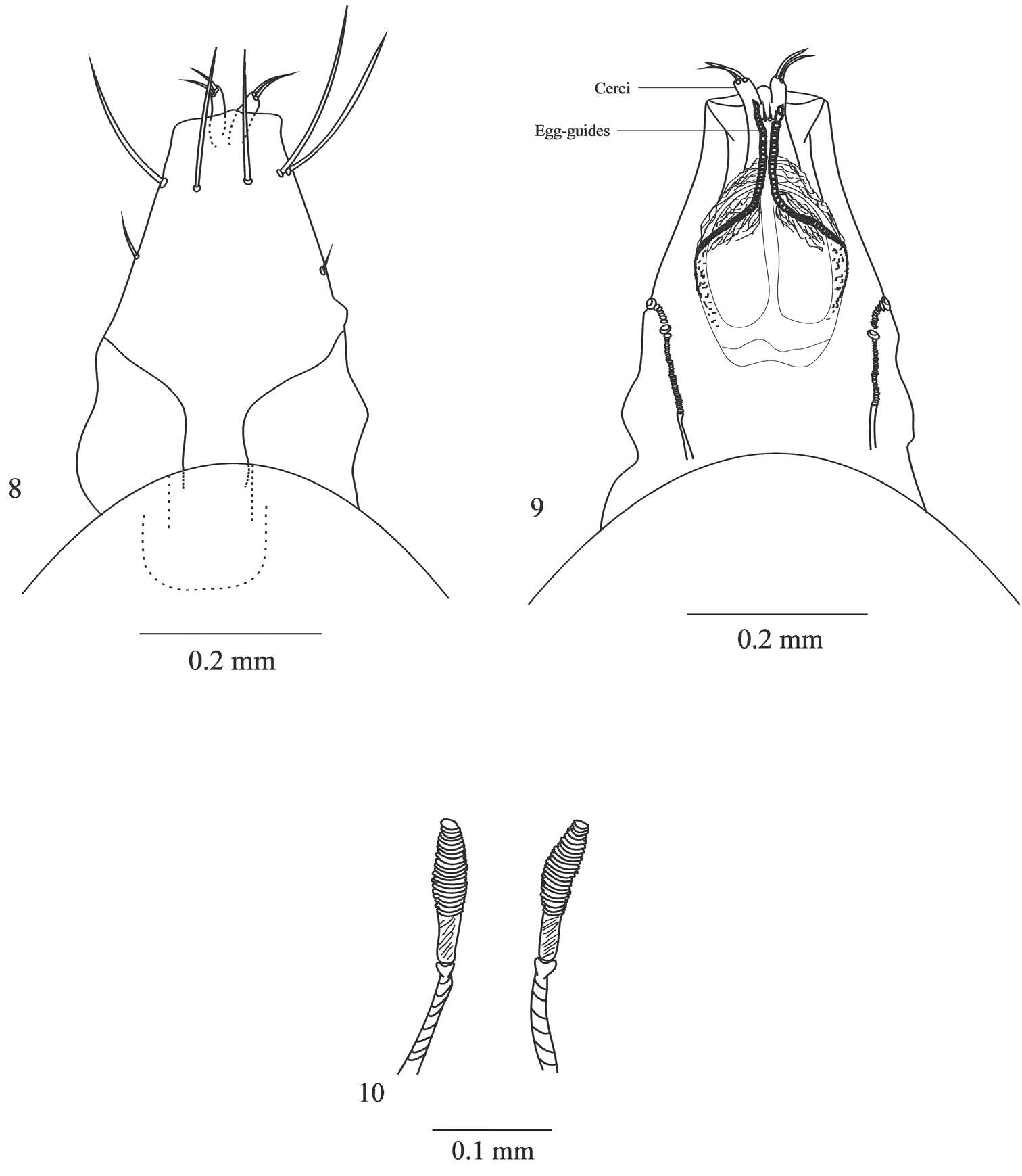
Viviane Rodrigues de Sousa, Márcia Souto Couri
Zookeys
Figures 8–10.Female ovipositor of Japanagromyza inferna Spencer 8 dorsal view 9 ventral view 10 spermathecae.
-
Shih-Tsai Yang, Hiromu Kurahashi, Shiuh-Feng Shiao
Zookeys
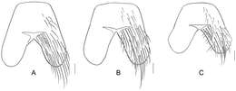
Figure 11.The male fifth abdominal sternites of Silbomyia species. A Silbomyia sauteri B Silbomyia hoeneana, Taiwan C Silbomyia hoeneana, China. Scale bars: 0.2 mm.
-
All Biocode files are based on field identifications to the best of the researcher’s ability at the time.