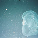
"During Dive 159 of the U.S. research submersible Deepstar 4000 on 22 October 1966 Dr Eric G. Barham, Dr George Pickwell, and Mr Ronald Church collected a remarkable scyphomedusan at a depth of about 723 m in the San Diego Trough. The specimen was collected by means of a suction device. In a letter to me Dr Barham said ' when first noted, the jellyfish's margin was collapsed and the exumbrella indented. As we maneuvered to bring the medusa in a position with the intake funnel of the collecting device, the vortex of the craft swirled the umbrella open. It was fully a metre in greatest dimensions '. 'Because of its size, not all of the specimen could fit into the pump container, and we returned to the surface with part of it still extending from the funnel.' Four of the photographs taken at the time are reproduced in Pl. I.
Dr Barham most kindly sent me the specimen for examination. He and his colleagues are to be congratulated on bringing this distinctive medusa successfully to the surface.
The medusa is incomplete. Most of the umbrella margin is missing as well as the central stomach region. All that came into the collecting device is however saved, including some portions of gonad and tissue with gastric cirri which had broken away from the body. Examination of the underwater photographs indicates that the medusa was in fact incomplete before it was caught. It was probably in a moribund state and was incapable of normal pulsation. The umbrella could be seen in some photographs to be distorted and twisted about by the turbulence caused by the Deepstar submersible. It appeared to be very tenuous and flexible.
In its preserved state the umbrella is obviously much contracted and probably compressed by the collecting apparatus. When spread out subumbrellar surface upwards it measured about 50 cm in diameter (Pl. II, fig. t). On opposite sides of the umbrella are two large tubular shaped processes. Dr Barham said that these were caused by bubbles of air which stretched the mesogloea. The umbrella has the form of a deep bell whose apex is missing. The mesogloea is solid and rather tough, but uniformly thin, being everywhere about 1-2 cm in thickness. It is much contracted and thrown into puckers and folds. It has a yellowish brown tinge and contains many mesogloeal cells. No epithelium is present. The mesogloea may have already been disintegrating and is of a floppy texture.
The radial canal system is most striking. It consists of a meshwork, likened by Dr Barham to wire-netting. The meshes are elongated radially and the whole system leads outwards to the umbrella margin. A portion of the umbrella showing the canal network is reproduced natural size in Pl. II, fig. 2. The whole network appears to be uniformly distributed over the umbrella and I could find no indications of any major straight canals leading from the centre to the periphery. The stomach portion of the umbrella is missing, but the network of canals can be seen to originate in the centre of the umbrella in a number of single straight canals of varying thickness which presumably connect with the stomach cavity.
In places around the margin there are portions of a narrow coronal muscle. Two such pieces can be seen in Pl. I, fig. 2, and they are also to be seen in Pl. II, fig. These portions of the margin are much contracted by the muscle and they gather the umbrella into little puckers. In its contracted state the muscle is about 15 mm in width.
The canal system at the margin in the region of the coronal muscle ends in a fine network of canals having the appearance shown in Text-fig. 1A. The meshes are compressed in a concertina-like fashion. A piece of stretched and mounted margin is shown in Text-fig. 1B. It is evident that there are complete but small meshes but it is not possible to decide whether they lead into a single ring canal. It is also not possible to determine whether there were any marginal tentacles.
The gonads are situated along the margins of fan-shaped mesenteries, and tend to be broken up into several isolated processes with incurved edges. The specimen is a male. The outer convex side of each process has thick mesogloea and the numerous oval sperm follicles are situated on the inner concave side embedded in the mesogloea (Text-fig. 2A). Each follicle is about 0.15— 0.25 mm in diameter and sections show spermatocytes and spermatozoa (Text-fig. 2 B).
Only one gonad was still attached to the umbrella. This was cut away for examination and photographing. It is shown natural size in Pl. III. The single straight canals which issue from the canal network in the central region can be seen running towards the base of the gonad mesentery. Some of these canals appear to be continued over the mesentery to the gonad tissue.
Three other gonads were found broken away. These had at the bases of their fan-shaped mesenteries an area of fragile tissue covered with many gastric cirri, possibly the subumbrella wall of the stomach. Where the fan-shaped gonadial mesentery narrows at its base it is continued into a grooved piece of solid mesogloea the sides of which have gastric cirri along them for a short distance. This solid mesogloea is continued as an elongated trough reminiscent of the axial groove of an oral arm. I am unable in fact to decipher what this could be. If it continued into the oral arm the gonads would be perradial, a position unknown in Scyphomedusae. In Text-fig. 3 I have drawn diagrammatically a gonad with this mesogloeal appendage and one is shown nearly natural size in Pl. IV. We know from the gonad shown in Pl. III that it is attached to the subumbrella. I have tried to suggest the situation in an inset sketch in Text-fig. 3, but until an intact specimen is obtained it is not possible to reconstruct the whole animal. Three of the appendages can be seen in Pl. I, fig. 4.
The gastric cirri had many nematocysts (Text-fig. 2c), but I could find none discharged. They were all of one kind, microbasic euryteles, 20-25 nm long and 8-11 nm wide. The gastric cirri were full of fat globules.
Until a more complete specimen of this medusa is obtained it is not possible to place it either in the Semaeostomeae or the Rhizostomeae, and its systematic position must remain uncertain. For this reason I name it Deepstaria enigmatica gen.nov., sp.nov., in recognition of its capture by Deepstar and its uncertain systematic position.
It is sufficient at this stage to regard the remarkable uniform network of the gastro-vascular canal system as a generic character. Such a canal system is not found in any known species of Scyphomedusae. It bears a resemblance, however, to the canal system of the ‘abnormal’ young medusae found in the viviparous species Stygiomedusa fabulosa (Russell, 1959; Russell & Rees, 1960, p. 313, Pl. IV), but it is perhaps stretching matters too far to suggest that this new species might be the sexual stage of the viviparous form.
The specimen had a large isopod in it, whose position is indicated in Pl. I, fig. I."
(Russell, 1967)